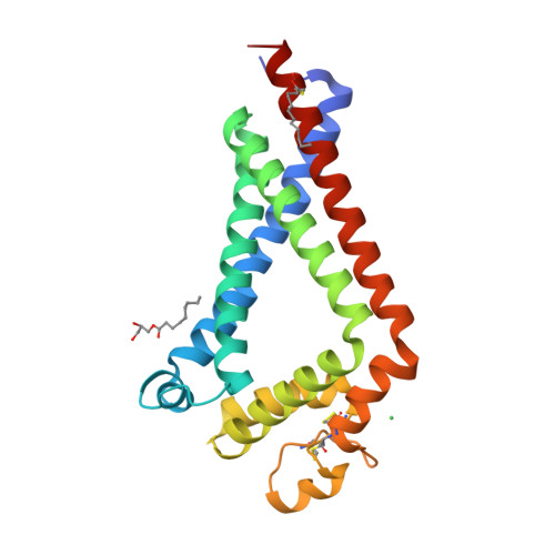
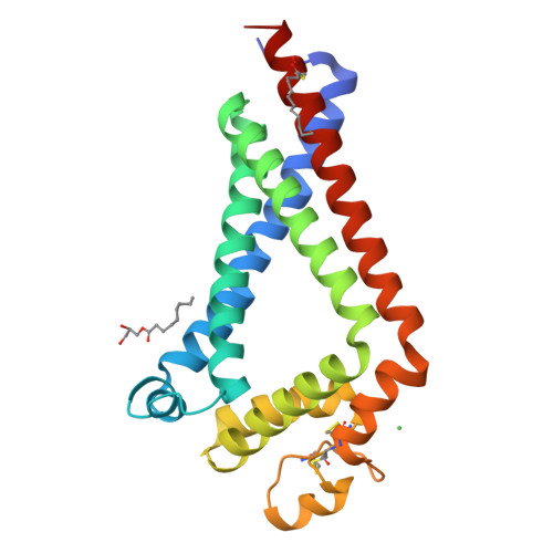
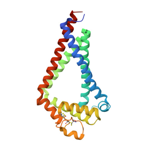
6K4J

Crystal Structure of the the CD9


Experimental Data Snapshot

  • Method: X-RAY DIFFRACTION
  • Resolution: 2.70 Å
  • R-Value Free: 0.289 
  • R-Value Work: 0.262 
  • R-Value Observed: 0.265 

wwPDB Validation   3D Report Full Report


Ligand Structure Quality Assessment 


This is version 1.1 of the entry. See complete history


Literature

Structural insights into tetraspanin CD9 function.

Umeda, R.Satouh, Y.Takemoto, M.Nakada-Nakura, Y.Liu, K.Yokoyama, T.Shirouzu, M.Iwata, S.Nomura, N.Sato, K.Ikawa, M.Nishizawa, T.Nureki, O.

(2020) Nat Commun 11: 1606-1606

  • DOI: https://doi.org/10.1038/s41467-020-15459-7
  • Primary Citation of Related Structures:  
    6K4J

  • PubMed Abstract: 

    Tetraspanins play critical roles in various physiological processes, ranging from cell adhesion to virus infection. The members of the tetraspanin family have four membrane-spanning domains and short and large extracellular loops, and associate with a broad range of other functional proteins to exert cellular functions. Here we report the crystal structure of CD9 and the cryo-electron microscopic structure of CD9 in complex with its single membrane-spanning partner protein, EWI-2. The reversed cone-like molecular shape of CD9 generates membrane curvature in the crystalline lipid layers, which explains the CD9 localization in regions with high membrane curvature and its implications in membrane remodeling. The molecular interaction between CD9 and EWI-2 is mainly mediated through the small residues in the transmembrane region and protein/lipid interactions, whereas the fertilization assay revealed the critical involvement of the LEL region in the sperm-egg fusion, indicating the different dependency of each binding domain for other partner proteins.


  • Organizational Affiliation

    Department of Biological Sciences Graduate School of Science, The University of Tokyo, 7-3-1 Hongo, Bunkyo-ku, Tokyo, Japan.


Macromolecules
Find similar proteins by:  (by identity cutoff)  |  3D Structure
Entity ID: 1
MoleculeChains Sequence LengthOrganismDetailsImage
CD9 antigen228Homo sapiensMutation(s): 0 
Gene Names: CD9MIC3TSPAN29GIG2
Membrane Entity: Yes 
UniProt & NIH Common Fund Data Resources
Find proteins for P21926 (Homo sapiens)
Explore P21926 
Go to UniProtKB:  P21926
PHAROS:  P21926
GTEx:  ENSG00000010278 
Entity Groups  
Sequence Clusters30% Identity50% Identity70% Identity90% Identity95% Identity100% Identity
UniProt GroupP21926
Sequence Annotations
Expand
  • Reference Sequence
Experimental Data & Validation

Experimental Data

  • Method: X-RAY DIFFRACTION
  • Resolution: 2.70 Å
  • R-Value Free: 0.289 
  • R-Value Work: 0.262 
  • R-Value Observed: 0.265 
  • Space Group: C 2 2 21
Unit Cell:
Length ( Å )Angle ( ˚ )
a = 45.18α = 90
b = 125.21β = 90
c = 129.4γ = 90
Software Package:
Software NamePurpose
XSCALEdata scaling
PHENIXrefinement
PDB_EXTRACTdata extraction
XDSdata reduction
PHASERphasing

Structure Validation

View Full Validation Report


Ligand Structure Quality Assessment 


Entry History 

Deposition Data

Revision History  (Full details and data files)

  • Version 1.0: 2020-05-13
    Type: Initial release
  • Version 1.1: 2024-11-06
    Changes: Data collection, Database references, Derived calculations, Structure summary