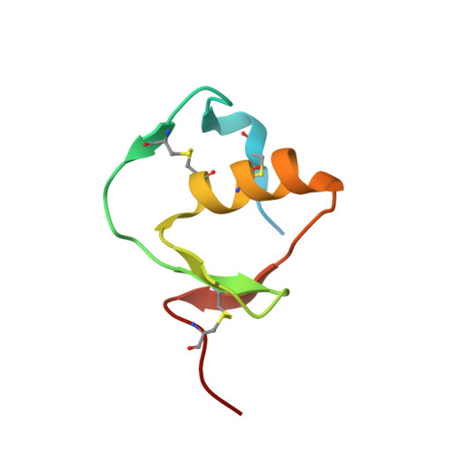
6KBR

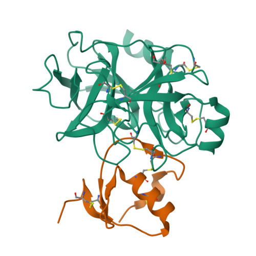
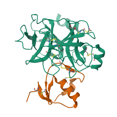
Crystal structure of Human KLK4 and SPINK2 derived KLK4 inhibitor complex


Experimental Data Snapshot

  • Method: X-RAY DIFFRACTION
  • Resolution: 2.00 Å
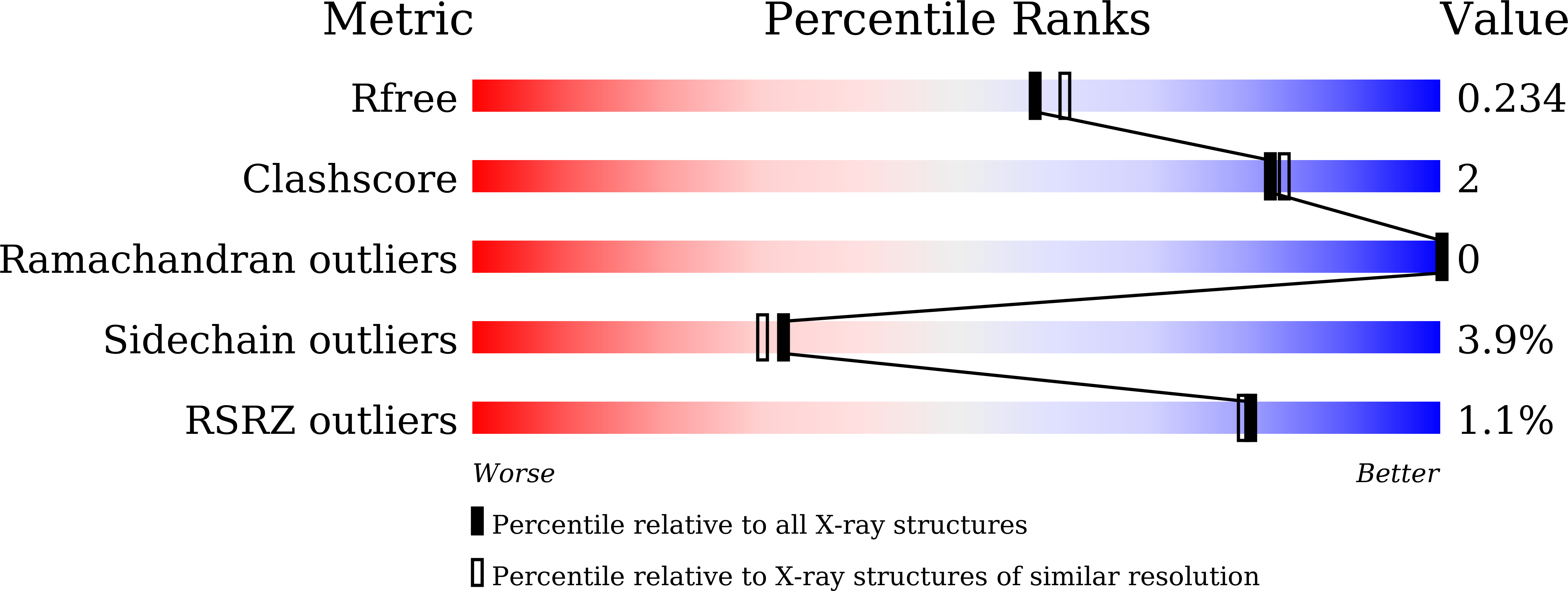
  • R-Value Free: 0.226 
  • R-Value Work: 0.185 
  • R-Value Observed: 0.187 

Starting Model: experimental
View more details

wwPDB Validation   3D Report Full Report


This is version 1.3 of the entry. See complete history


Literature

A protein scaffold, engineered SPINK2, for generation of inhibitors with high affinity and specificity against target proteases.

Nishimiya, D.Kawaguchi, Y.Kodama, S.Nasu, H.Yano, H.Yamaguchi, A.Tamura, M.Hashimoto, R.

(2019) Sci Rep 9: 11436-11436

  • DOI: https://doi.org/10.1038/s41598-019-47615-5
  • Primary Citation of Related Structures:  
    6KBR

  • PubMed Abstract: 

    Proteases are one of attractive therapeutic targets to play key roles in pharmacological action. There are many protease inhibitors in nature, and most of them structurally have cystine knot motifs. Their structures are favorable for recognition of active pockets of proteases, leading to the potent inhibition. However, they also have drawbacks, such as broad cross-reactivity, on the therapeutic application. To create therapeutic proteins derived from a disulfide-rich scaffold, we selected human serine protease inhibitor Kazal type 2 (SPINK2) through a scaffold screening, as a protein scaffold with requirements for therapeutic proteins. We then constructed a diverse library of the engineered SPINK2 by introducing random mutations into its flexible loop region with the designed method. By phage panning against four serine proteases, we isolated potent inhibitors against each target with picomolar K D and sub-nanomolar K i values. Also, they exhibited the desired specificities against target proteases without inhibiting non-target proteases. The crystal structure of kallikrein related peptidase 4 (KLK4)-engineered SPINK2 complex revealed the interface with extensive conformational complementarity. Our study demonstrates that engineered SPINK2 can serve as a scaffold to generate therapeutic molecules against target proteins with groove structures.


  • Organizational Affiliation

    DAIICHI SANKYO CO., LTD., Biologics Division, Modality Research Laboratories, 1-2-58, Hiromachi, Shinagawa-ku, Tokyo, 140-8710, Japan. nishimiya.daisuke.tc@daiichisankyo.co.jp.


Macromolecules
Find similar proteins by:  (by identity cutoff)  |  3D Structure
Entity ID: 1
MoleculeChains Sequence LengthOrganismDetailsImage
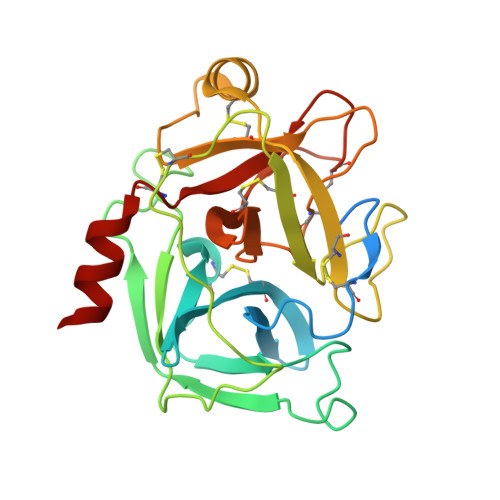
Kallikrein-4254Homo sapiensMutation(s): 0 
Gene Names: KLK4EMSP1PRSS17PSTS
EC: 3.4.21
UniProt & NIH Common Fund Data Resources
Find proteins for Q9Y5K2 (Homo sapiens)
Explore Q9Y5K2 
Go to UniProtKB:  Q9Y5K2
PHAROS:  Q9Y5K2
GTEx:  ENSG00000167749 
Entity Groups  
Sequence Clusters30% Identity50% Identity70% Identity90% Identity95% Identity100% Identity
UniProt GroupQ9Y5K2
Sequence Annotations
Expand
  • Reference Sequence
Find similar proteins by:  (by identity cutoff)  |  3D Structure
Entity ID: 2
MoleculeChains Sequence LengthOrganismDetailsImage
K41043B [auth C]65Homo sapiensMutation(s): 0 
UniProt & NIH Common Fund Data Resources
Find proteins for P20155 (Homo sapiens)
Explore P20155 
Go to UniProtKB:  P20155
PHAROS:  P20155
GTEx:  ENSG00000128040 
Entity Groups  
Sequence Clusters30% Identity50% Identity70% Identity90% Identity95% Identity100% Identity
UniProt GroupP20155
Sequence Annotations
Expand
  • Reference Sequence
Small Molecules
Ligands 1 Unique
IDChains Name / Formula / InChI Key2D Diagram3D Interactions
GOL
Query on GOL

Download Ideal Coordinates CCD File 
C [auth A]GLYCEROL
C3 H8 O3
PEDCQBHIVMGVHV-UHFFFAOYSA-N
Experimental Data & Validation

Experimental Data

  • Method: X-RAY DIFFRACTION
  • Resolution: 2.00 Å
  • R-Value Free: 0.226 
  • R-Value Work: 0.185 
  • R-Value Observed: 0.187 
  • Space Group: C 1 2 1
Unit Cell:
Length ( Å )Angle ( ˚ )
a = 145.208α = 90
b = 41.62β = 91.58
c = 43.59γ = 90
Software Package:
Software NamePurpose
REFMACrefinement
iMOSFLMdata reduction
MOLREPphasing

Structure Validation

View Full Validation Report


Entry History 

Deposition Data

Revision History  (Full details and data files)

  • Version 1.0: 2019-07-17
    Type: Initial release
  • Version 1.1: 2019-08-21
    Changes: Data collection, Database references
  • Version 1.2: 2023-11-22
    Changes: Data collection, Database references, Refinement description
  • Version 1.3: 2024-10-23
    Changes: Structure summary