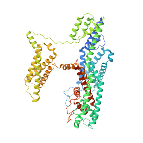
6NQ2

Cryo-EM structure of human TPC2 channel in the ligand-bound closed state


Experimental Data Snapshot

  • Method: ELECTRON MICROSCOPY
  • Resolution: 3.40 Å
  • Aggregation State: PARTICLE 
  • Reconstruction Method: SINGLE PARTICLE 

wwPDB Validation   3D Report Full Report


This is version 1.1 of the entry. See complete history


Literature

Structural mechanisms of phospholipid activation of the human TPC2 channel.

She, J.Zeng, W.Guo, J.Chen, Q.Bai, X.Jiang, Y.

(2019) Elife 8

  • DOI: https://doi.org/10.7554/eLife.45222
  • Primary Citation of Related Structures:  
    6NQ0, 6NQ1, 6NQ2

  • PubMed Abstract: 

    Mammalian two-pore channels (TPCs) regulate the physiological functions of the endolysosome. Here we present cryo-EM structures of human TPC2 (HsTPC2), a phosphatidylinositol 3,5-bisphosphate (PI(3,5)P 2 )-activated, Na + selective channel, in the ligand-bound and apo states. The apo structure captures the closed conformation, while the ligand-bound form features the channel in both open and closed conformations. Combined with functional analysis, these structures provide insights into the mechanism of PI(3,5)P 2 -regulated gating of TPC2, which is distinct from that of TPC1. Specifically, the endolysosome-specific PI(3,5)P 2 binds at the first 6-TM and activates the channel - independently of the membrane potential - by inducing a structural change at the pore-lining inner helix (IS6), which forms a continuous helix in the open state but breaks into two segments at Gly317 in the closed state. Additionally, structural comparison to the voltage-dependent TPC1 structure allowed us to identify Ile551 as being responsible for the loss of voltage dependence in TPC2.


  • Organizational Affiliation

    Department of Physiology, University of Texas Southwestern Medical Center, Dallas, United States.


Macromolecules
Find similar proteins by:  (by identity cutoff)  |  3D Structure
Entity ID: 1
MoleculeChains Sequence LengthOrganismDetailsImage
Two pore calcium channel protein 2
A, B
756Homo sapiensMutation(s): 0 
Gene Names: TPCN2TPC2
Membrane Entity: Yes 
UniProt & NIH Common Fund Data Resources
Find proteins for Q8NHX9 (Homo sapiens)
Explore Q8NHX9 
Go to UniProtKB:  Q8NHX9
PHAROS:  Q8NHX9
GTEx:  ENSG00000162341 
Entity Groups  
Sequence Clusters30% Identity50% Identity70% Identity90% Identity95% Identity100% Identity
UniProt GroupQ8NHX9
Sequence Annotations
Expand
  • Reference Sequence
Small Molecules
Ligands 1 Unique
IDChains Name / Formula / InChI Key2D Diagram3D Interactions
EUJ
Query on EUJ

Download Ideal Coordinates CCD File 
C [auth A],
D [auth B]
(2R)-3-{[(S)-hydroxy{[(1S,2R,3R,4S,5S,6R)-2,4,6-trihydroxy-3,5-bis(phosphonooxy)cyclohexyl]oxy}phosphoryl]oxy}propane-1,2-diyl dioctanoate
C25 H49 O19 P3
QXHVLVSULWMTCV-DICZBTHZSA-N
Experimental Data & Validation

Experimental Data

  • Method: ELECTRON MICROSCOPY
  • Resolution: 3.40 Å
  • Aggregation State: PARTICLE 
  • Reconstruction Method: SINGLE PARTICLE 

Structure Validation

View Full Validation Report


Entry History & Funding Information

Deposition Data


Funding OrganizationLocationGrant Number
National Institutes of Health/National Institute of General Medical Sciences (NIH/NIGMS)United StatesGM079179
Howard Hughes Medical Institute (HHMI)United States--
Welch FoundationGrant I-1578
Cancer Prevention and Research Institute of Texas (CPRIT)United States--

Revision History  (Full details and data files)

  • Version 1.0: 2019-03-27
    Type: Initial release
  • Version 1.1: 2019-11-20
    Changes: Author supporting evidence