6O1G | pdb_00006o1g

Full length human plasma kallikrein with inhibitor


Experimental Data Snapshot

  • Method: X-RAY DIFFRACTION
  • Resolution: 2.20 Å
  • R-Value Free: 
    0.240 (Depositor), 0.242 (DCC) 
  • R-Value Work: 
    0.201 (Depositor), 0.205 (DCC) 
  • R-Value Observed: 
    0.203 (Depositor) 

wwPDB Validation   3D Report Full Report


Ligand Structure Quality Assessment 

Created with Raphaël 2.3.0Worse 01 BetterLigand structure goodness of fit to experimental dataBest fitted 7SDClick on this verticalbar to view details

This is version 1.4 of the entry. See complete history


Literature

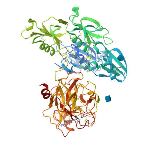
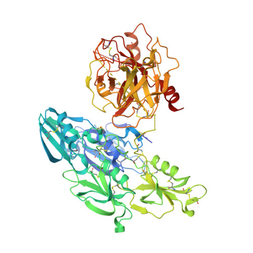
Structures of full-length plasma kallikrein bound to highly specific inhibitors describe a new mode of targeted inhibition.

Partridge, J.R.Choy, R.M.Silva-Garcia, A.Yu, C.Li, Z.Sham, H.Metcalf, B.

(2019) J Struct Biol 206: 170-182

  • DOI: https://doi.org/10.1016/j.jsb.2019.03.001
  • Primary Citation of Related Structures:  
    6BFP, 6O1G, 6O1S

  • PubMed Abstract: 

    Plasma kallikrein (pKal) is a serine protease responsible for cleaving high-molecular-weight kininogen to produce the pro-inflammatory peptide, bradykinin. Unregulated pKal activity can lead to hereditary angioedema (HAE) following excess bradykinin release. HAE attacks can lead to a compromised airway that can be life threatening. As there are limited agents for prophylaxis of HAE attacks, there is a high unmet need for a therapeutic agent for regulating pKal with a high degree of specificity. Here we present crystal structures of both full-length and the protease domain of pKal, bound to two very distinct classes of small-molecule inhibitors: compound 1, and BCX4161. Both inhibitors demonstrate low nM inhibitory potency for pKal and varying specificity for related serine proteases. Compound 1 utilizes a surprising mode of interaction and upon binding results in a rearrangement of the binding pocket. Co-crystal structures of pKal describes why this class of small-molecule inhibitor is potent. Lack of conservation in surrounding residues explains the ∼10,000-fold specificity over structurally similar proteases, as shown by in vitro protease inhibition data. Structural information, combined with biochemical and enzymatic analyses, provides a novel scaffold for the design of targeted oral small molecule inhibitors of pKal for treatment of HAE and other diseases resulting from unregulated plasma kallikrein activity.


  • Organizational Affiliation

    Global Blood Therapeutics, South San Francisco, CA 94080, United States. Electronic address: jpartridge@gbt.com.


Macromolecules
Find similar proteins by:  (by identity cutoff)  |  3D Structure
Entity ID: 1
MoleculeChains Sequence LengthOrganismDetailsImage
Plasma kallikrein638Homo sapiensMutation(s): 0 
EC: 3.4.21.34
UniProt & NIH Common Fund Data Resources
Find proteins for P03952 (Homo sapiens)
Explore P03952 
Go to UniProtKB:  P03952
PHAROS:  P03952
GTEx:  ENSG00000164344 
Entity Groups  
Sequence Clusters30% Identity50% Identity70% Identity90% Identity95% Identity100% Identity
UniProt GroupP03952
Sequence Annotations
Expand
  • Reference Sequence
Small Molecules
Binding Affinity Annotations 
IDSourceBinding Affinity
7SD BindingDB:  6O1G IC50: min: 0.2, max: 2.4 (nM) from 2 assay(s)
Experimental Data & Validation

Experimental Data

  • Method: X-RAY DIFFRACTION
  • Resolution: 2.20 Å
  • R-Value Free:  0.240 (Depositor), 0.242 (DCC) 
  • R-Value Work:  0.201 (Depositor), 0.205 (DCC) 
  • R-Value Observed: 0.203 (Depositor) 
Space Group: P 1 21 1
Unit Cell:
Length ( Å )Angle ( ˚ )
a = 55.97α = 90
b = 121.92β = 105.79
c = 65.89γ = 90
Software Package:
Software NamePurpose
PHENIXrefinement
iMOSFLMdata reduction
SCALAdata scaling
PHASERphasing

Structure Validation

View Full Validation Report


Ligand Structure Quality Assessment 

Created with Raphaël 2.3.0Worse 01 BetterLigand structure goodness of fit to experimental dataBest fitted 7SDClick on this verticalbar to view details

Entry History 

Deposition Data

Revision History  (Full details and data files)

  • Version 1.0: 2019-03-06
    Type: Initial release
  • Version 1.1: 2019-03-27
    Changes: Data collection, Database references
  • Version 1.2: 2019-05-15
    Changes: Data collection, Database references
  • Version 1.3: 2020-07-29
    Type: Remediation
    Reason: Carbohydrate remediation
    Changes: Data collection, Derived calculations, Structure summary
  • Version 1.4: 2024-10-16
    Changes: Data collection, Database references, Structure summary