6P0P

Human beta-tryptase co-crystal structure with 5-{4-[3-(aminomethyl)phenyl]piperidine-1-carbonyl}-2-(3'-{4-[3-(aminomethyl)phenyl]piperidine-1-carbonyl}-[1,1'-biphenyl]-3-yl)-2-hydroxy-2H-1,3,2-benzodioxaborol-2-uide


Experimental Data Snapshot

  • Method: X-RAY DIFFRACTION
  • Resolution: 2.55 Å
  • R-Value Free: 0.249 
  • R-Value Work: 0.180 
  • R-Value Observed: 0.184 

wwPDB Validation   3D Report Full Report


Ligand Structure Quality Assessment 


This is version 1.2 of the entry. See complete history


Literature

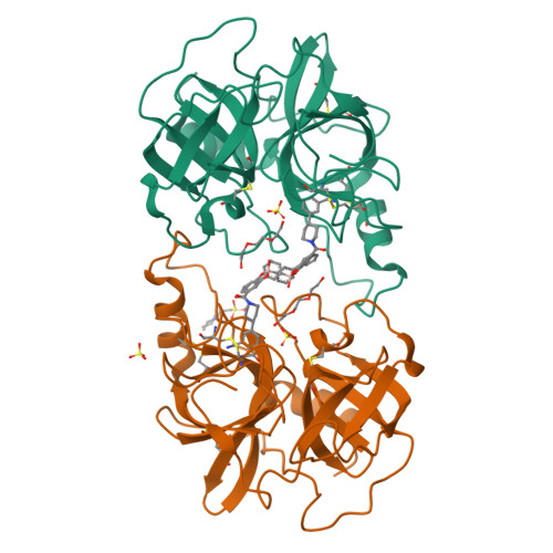
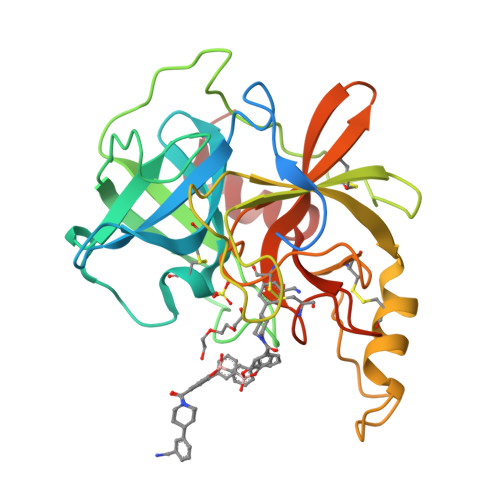
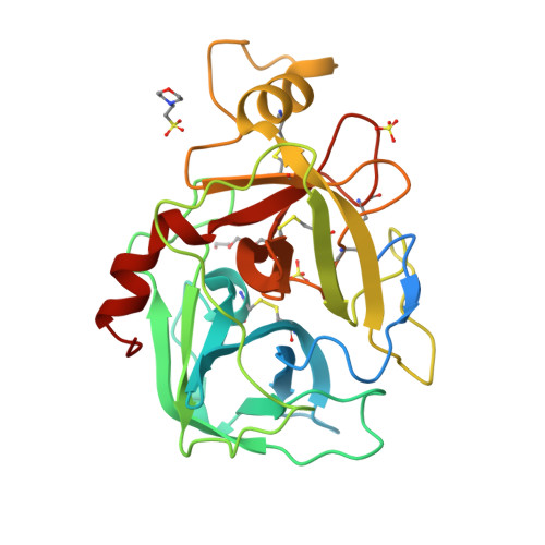
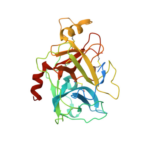
Novel, Self-Assembling Dimeric Inhibitors of Human beta Tryptase.

Giardina, S.F.Werner, D.S.Pingle, M.Feinberg, P.B.Foreman, K.W.Bergstrom, D.E.Arnold, L.D.Barany, F.

(2020) J Med Chem 63: 3004-3027

  • DOI: https://doi.org/10.1021/acs.jmedchem.9b01689
  • Primary Citation of Related Structures:  
    6P0P

  • PubMed Abstract: 

    β-Tryptase, a homotetrameric serine protease, has four identical active sites facing a central pore, presenting an optimized setting for the rational design of bivalent inhibitors that bridge two adjacent sites. Using diol, hydroxymethyl phenols or benzoyl methyl hydroxamates, and boronic acid chemistries to reversibly join two [3-(1-acylpiperidin-4-yl)phenyl]methanamine core ligands, we have successfully produced a series of self-assembling heterodimeric inhibitors. These heterodimeric tryptase inhibitors demonstrate superior activity compared to monomeric modes of inhibition. X-ray crystallography validated the dimeric mechanism of inhibition, and compounds demonstrated high selectivity against related proteases, good target engagement, and tryptase inhibition in HMC1 xenograft models. Screening 3872 possible combinations from 44 boronic acid and 88 diol derivatives revealed several combinations that produced nanomolar inhibition, and seven unique pairs produced greater than 100-fold improvement in potency over monomeric inhibition. These heterodimeric tryptase inhibitors demonstrate the power of target-driven combinatorial chemistry to deliver bivalent drugs in a small molecule form.


  • Organizational Affiliation

    Department of Microbiology and Immunology, Weill Cornell Medicine, 1300 York Avenue, Box 62, New York, New York 10065, United States.


Macromolecules
Find similar proteins by:  (by identity cutoff)  |  3D Structure
Entity ID: 1
MoleculeChains Sequence LengthOrganismDetailsImage
Tryptase alpha/beta-1
A, B
275Homo sapiensMutation(s): 0 
Gene Names: TPSAB1TPS1TPS2TPSB1
EC: 3.4.21.59
UniProt & NIH Common Fund Data Resources
Find proteins for Q15661 (Homo sapiens)
Explore Q15661 
Go to UniProtKB:  Q15661
PHAROS:  Q15661
GTEx:  ENSG00000172236 
Entity Groups  
Sequence Clusters30% Identity50% Identity70% Identity90% Identity95% Identity100% Identity
UniProt GroupQ15661
Sequence Annotations
Expand
  • Reference Sequence
Small Molecules
Ligands 4 Unique
IDChains Name / Formula / InChI Key2D Diagram3D Interactions
Y35 (Subject of Investigation/LOI)
Query on Y35

Download Ideal Coordinates CCD File 
C [auth A](3'-{4-[3-(aminomethyl)phenyl]piperidine-1-carbonyl}[1,1'-biphenyl]-3-yl){4-[3-(aminomethyl)phenyl]piperidin-1-yl}[3,4-di(hydroxy-kappaO)phenyl]methanonato(2-)hydroxyborate(1-)
C44 H46 B N4 O5
BXWXLCXUQLTPFH-WBVITSLISA-N
MES
Query on MES

Download Ideal Coordinates CCD File 
H [auth B]2-(N-MORPHOLINO)-ETHANESULFONIC ACID
C6 H13 N O4 S
SXGZJKUKBWWHRA-UHFFFAOYSA-N
PGE
Query on PGE

Download Ideal Coordinates CCD File 
E [auth A],
I [auth B]
TRIETHYLENE GLYCOL
C6 H14 O4
ZIBGPFATKBEMQZ-UHFFFAOYSA-N
SO4
Query on SO4

Download Ideal Coordinates CCD File 
D [auth A],
F [auth B],
G [auth B]
SULFATE ION
O4 S
QAOWNCQODCNURD-UHFFFAOYSA-L
Experimental Data & Validation

Experimental Data

  • Method: X-RAY DIFFRACTION
  • Resolution: 2.55 Å
  • R-Value Free: 0.249 
  • R-Value Work: 0.180 
  • R-Value Observed: 0.184 
  • Space Group: P 31 2 1
Unit Cell:
Length ( Å )Angle ( ˚ )
a = 78.099α = 90
b = 78.099β = 90
c = 165.182γ = 120
Software Package:
Software NamePurpose
REFMACrefinement
HKL-2000data reduction
SCALEPACKdata scaling
MOLREPphasing

Structure Validation

View Full Validation Report


Ligand Structure Quality Assessment 


Entry History 

Deposition Data

Revision History  (Full details and data files)

  • Version 1.0: 2020-03-25
    Type: Initial release
  • Version 1.1: 2020-04-08
    Changes: Database references
  • Version 1.2: 2023-10-11
    Changes: Data collection, Database references, Refinement description