6PXW

Cryo-EM structure of full-length insulin receptor bound to 4 insulin. 3D refinement was focused on the top part of the receptor complex.


Experimental Data Snapshot

  • Method: ELECTRON MICROSCOPY
  • Resolution: 3.10 Å
  • Aggregation State: PARTICLE 
  • Reconstruction Method: SINGLE PARTICLE 

wwPDB Validation   3D Report Full Report


This is version 1.0 of the entry. See complete history


Literature

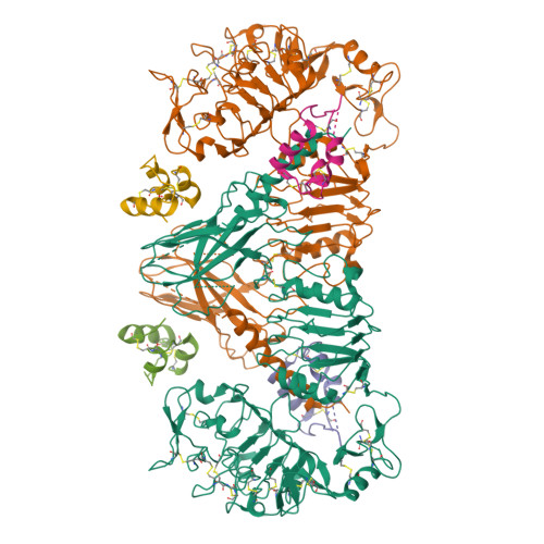
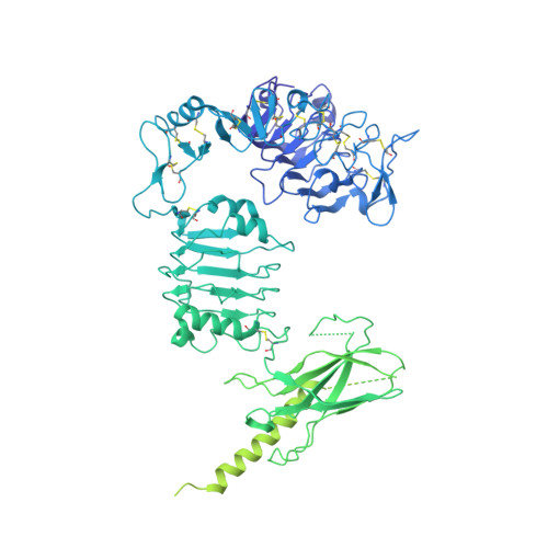
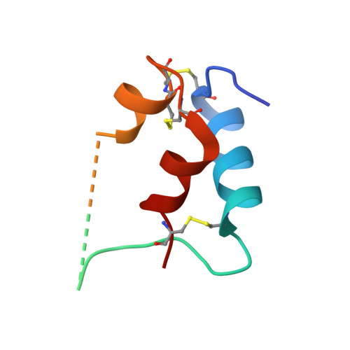
Activation mechanism of the insulin receptor revealed by cryo-EM structure of the fully liganded receptor-ligand complex.

Uchikawa, E.Choi, E.Shang, G.Yu, H.Bai, X.C.

(2019) Elife 8

  • DOI: https://doi.org/10.7554/eLife.48630
  • Primary Citation of Related Structures:  
    6PXV, 6PXW

  • PubMed Abstract: 

    Insulin signaling controls metabolic homeostasis. Here, we report the cryo-EM structure of full-length insulin receptor (IR) and insulin complex in the active state. This structure unexpectedly reveals that maximally four insulins can bind the 'T'-shaped IR dimer at four distinct sites related by 2-fold symmetry. Insulins 1 and 1' bind to sites 1 and 1', formed by L1 of one IR protomer and α-CT and FnIII-1 of the other. Insulins 2 and 2' bind to sites 2 and 2' on FnIII-1 of each protomer. Mutagenesis and cellular assays show that both sites 1 and 2 are required for optimal insulin binding and IR activation. We further identify a homotypic FnIII-2-FnIII-2 interaction in mediating the dimerization of membrane proximal domains in the active IR dimer. Our results indicate that binding of multiple insulins at two distinct types of sites disrupts the autoinhibited apo-IR dimer and stabilizes the active dimer.


  • Organizational Affiliation

    Department of Biophysics, University of Texas Southwestern Medical Center, Dallas, United States.


Macromolecules
Find similar proteins by:  (by identity cutoff)  |  3D Structure
Entity ID: 1
MoleculeChains Sequence LengthOrganismDetailsImage
Insulin receptor
A, B
1,354Homo sapiensMutation(s): 0 
Gene Names: INSR
EC: 2.7.10.1
UniProt & NIH Common Fund Data Resources
Find proteins for P06213 (Homo sapiens)
Explore P06213 
Go to UniProtKB:  P06213
PHAROS:  P06213
GTEx:  ENSG00000171105 
Entity Groups  
Sequence Clusters30% Identity50% Identity70% Identity90% Identity95% Identity100% Identity
UniProt GroupP06213
Sequence Annotations
Expand
  • Reference Sequence
Find similar proteins by:  (by identity cutoff)  |  3D Structure
Entity ID: 2
MoleculeChains Sequence LengthOrganismDetailsImage
Insulin
C, D, E, F
74Homo sapiensMutation(s): 0 
Gene Names: INS
UniProt & NIH Common Fund Data Resources
Find proteins for A6XGL2 (Homo sapiens)
Explore A6XGL2 
Go to UniProtKB:  A6XGL2
GTEx:  ENSG00000254647 
Entity Groups  
Sequence Clusters30% Identity50% Identity70% Identity90% Identity95% Identity100% Identity
UniProt GroupA6XGL2
Sequence Annotations
Expand
  • Reference Sequence
Experimental Data & Validation

Experimental Data

  • Method: ELECTRON MICROSCOPY
  • Resolution: 3.10 Å
  • Aggregation State: PARTICLE 
  • Reconstruction Method: SINGLE PARTICLE 
EM Software:
TaskSoftware PackageVersion
MODEL REFINEMENTPHENIX
RECONSTRUCTIONRELION

Structure Validation

View Full Validation Report


Entry History 

Deposition Data

Revision History  (Full details and data files)

  • Version 1.0: 2019-09-04
    Type: Initial release