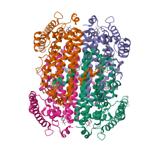
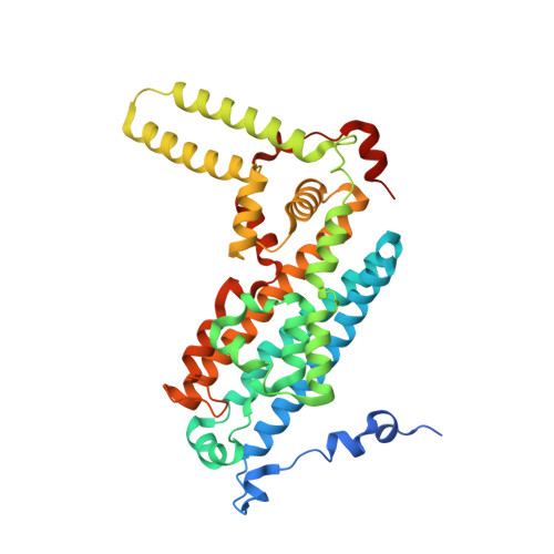
6PYZ

Crystal Structure of human Tryptophan 2,3-dioxygenase in complex with PF-06840003 in Active Site


Experimental Data Snapshot

  • Method: X-RAY DIFFRACTION
  • Resolution: 2.02 Å
  • R-Value Free: 0.243 
  • R-Value Work: 0.189 
  • R-Value Observed: 0.192 

Starting Model: experimental
View more details

wwPDB Validation   3D Report Full Report


Ligand Structure Quality Assessment 


This is version 2.0 of the entry. See complete history


Literature

Structural Basis of Inhibitor Selectivity in Human Indoleamine 2,3-Dioxygenase 1 and Tryptophan Dioxygenase.

Pham, K.N.Lewis-Ballester, A.Yeh, S.R.

(2019) J Am Chem Soc 141: 18771-18779

  • DOI: https://doi.org/10.1021/jacs.9b08871
  • Primary Citation of Related Structures:  
    6PYY, 6PYZ, 6PZ1

  • PubMed Abstract: 

    Indoleamine 2,3-dioxygenase 1 (hIDO1) and tryptophan dioxygenase (hTDO) are two of the only three heme-based dioxygenases in humans. They have recently been identified as key cancer immunotherapeutic drug targets. While structures of hIDO1 in complex with inhibitors have been documented, so far there are no structures of hTDO-inhibitor complexes available. Here we use PF-06840003 (IPD), a hIDO1-selective inhibitor in clinical trials, as a structural probe to elucidate inhibitor-selectivity in hIDO1 versus hTDO. Spectroscopic studies show that IPD exhibits 400-fold higher inhibition activity toward hIDO1 with respect to hTDO. Crystallographic structures reveal that the binding pocket of IPD in the active site in hIDO1 is much more flexible as compared to that in hTDO, which offers a molecular explanation for the superior inhibition activity of IPD in hIDO1 with respect to hTDO. In addition to the IPD bound in the active site, a second IPD molecule was identified in an inhibitory site on the proximal side of the heme in hIDO1 and in an exosite that is ∼40 Å away from the active site in hTDO. Taken together the data provide new insights into structure-based design of mono and dual inhibitors targeting hIDO1 and/or hTDO.


  • Organizational Affiliation

    Department of Physiology and Biophysics , Albert Einstein College of Medicine , The Bronx , New York 10461 , United States.


Macromolecules
Find similar proteins by:  (by identity cutoff)  |  3D Structure
Entity ID: 1
MoleculeChains Sequence LengthOrganismDetailsImage
Tryptophan 2,3-dioxygenase
A, B, C, D
380Homo sapiensMutation(s): 0 
Gene Names: TDO2TDO
EC: 1.13.11.11
UniProt & NIH Common Fund Data Resources
Find proteins for P48775 (Homo sapiens)
Explore P48775 
Go to UniProtKB:  P48775
PHAROS:  P48775
GTEx:  ENSG00000151790 
Entity Groups  
Sequence Clusters30% Identity50% Identity70% Identity90% Identity95% Identity100% Identity
UniProt GroupP48775
Sequence Annotations
Expand
  • Reference Sequence
Small Molecules
Ligands 3 Unique
IDChains Name / Formula / InChI Key2D Diagram3D Interactions
HEM (Subject of Investigation/LOI)
Query on HEM

Download Ideal Coordinates CCD File 
E [auth A],
H [auth B],
K [auth C],
N [auth D]
PROTOPORPHYRIN IX CONTAINING FE
C34 H32 Fe N4 O4
KABFMIBPWCXCRK-RGGAHWMASA-L
H7S (Subject of Investigation/LOI)
Query on H7S

Download Ideal Coordinates CCD File 
F [auth A],
I [auth B],
L [auth C],
O [auth D]
(3S)-3-(5-fluoro-1H-indol-3-yl)pyrrolidine-2,5-dione
C12 H9 F N2 O2
MXKLDYKORJEOPR-QMMMGPOBSA-N
ZIQ (Subject of Investigation/LOI)
Query on ZIQ

Download Ideal Coordinates CCD File 
G [auth A],
J [auth B],
M [auth C],
P [auth D]
alpha-methyl-L-tryptophan
C12 H14 N2 O2
ZTTWHZHBPDYSQB-LBPRGKRZSA-N
Binding Affinity Annotations 
IDSourceBinding Affinity
H7S BindingDB:  6PYZ IC50: min: 5.00e+4, max: 5.00e+4 (nM) from 2 assay(s)
Experimental Data & Validation

Experimental Data

  • Method: X-RAY DIFFRACTION
  • Resolution: 2.02 Å
  • R-Value Free: 0.243 
  • R-Value Work: 0.189 
  • R-Value Observed: 0.192 
  • Space Group: P 21 21 2
Unit Cell:
Length ( Å )Angle ( ˚ )
a = 143.648α = 90
b = 154.158β = 90
c = 87.951γ = 90
Software Package:
Software NamePurpose
REFMACrefinement
PDB_EXTRACTdata extraction
XDSdata reduction
SCALAdata scaling
PHASERphasing

Structure Validation

View Full Validation Report


Ligand Structure Quality Assessment 


Entry History & Funding Information

Deposition Data


Funding OrganizationLocationGrant Number
National Institutes of Health/National Institute of General Medical Sciences (NIH/NIGMS)United StatesGM115773
National Science Foundation (NSF, United States)United StatesCHE-1404929

Revision History  (Full details and data files)

  • Version 1.0: 2020-01-01
    Type: Initial release
  • Version 1.1: 2023-10-11
    Changes: Data collection, Database references, Derived calculations, Refinement description
  • Version 2.0: 2023-11-15
    Changes: Atomic model, Data collection