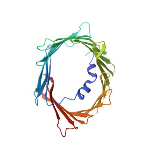
6TIR

NOE based model of hVDAC-1 bound to beta-NADH in detergent micelles


Experimental Data Snapshot

  • Method: SOLUTION NMR
  • Conformers Calculated: 500 
  • Conformers Submitted: 10 
  • Selection Criteria: structures with the lowest energy 

wwPDB Validation   3D Report Full Report


This is version 1.3 of the entry. See complete history


Literature

The Structural Basis for Low Conductance in the Membrane Protein VDAC upon beta-NADH Binding and Voltage Gating.

Bohm, R.Amodeo, G.F.Murlidaran, S.Chavali, S.Wagner, G.Winterhalter, M.Brannigan, G.Hiller, S.

(2020) Structure 28: 206-214.e4

  • DOI: https://doi.org/10.1016/j.str.2019.11.015
  • Primary Citation of Related Structures:  
    6TIQ, 6TIR

  • PubMed Abstract: 

    The voltage-dependent anion channel (VDAC) forms the primary diffusion pore of the outer mitochondrial membrane. In its apo form, VDAC adopts an open conformation with high conductance. States of lower conductance can be induced by ligand binding or the application of voltage. Here, we clarify at the atomic level how β-NADH binding leads to a low-conductance state and characterize the role of the VDAC N-terminal helix in voltage gating. A high-resolution NMR structure of human VDAC-1 with bound NADH, combined with molecular dynamics simulation show that β-NADH binding reduces the pore conductance sterically without triggering a structural change. Electrophysiology recordings of crosslinked protein variants and NMR relaxation experiments probing different time scales show that increased helix dynamics is present in the open state and that motions of the N-terminal helices are involved in the VDAC voltage gating mechanism.


  • Organizational Affiliation

    Biozentrum, University of Basel, Basel 4056, Switzerland.


Macromolecules
Find similar proteins by:  (by identity cutoff)  |  3D Structure
Entity ID: 1
MoleculeChains Sequence LengthOrganismDetailsImage
Voltage-dependent anion-selective channel protein 1291Homo sapiensMutation(s): 0 
Gene Names: VDAC1VDAC
Membrane Entity: Yes 
UniProt & NIH Common Fund Data Resources
Find proteins for P21796 (Homo sapiens)
Explore P21796 
Go to UniProtKB:  P21796
PHAROS:  P21796
GTEx:  ENSG00000213585 
Entity Groups  
Sequence Clusters30% Identity50% Identity70% Identity90% Identity95% Identity100% Identity
UniProt GroupP21796
Sequence Annotations
Expand
  • Reference Sequence
Small Molecules
Ligands 1 Unique
IDChains Name / Formula / InChI Key2D Diagram3D Interactions
NAI (Subject of Investigation/LOI)
Query on NAI

Download Ideal Coordinates CCD File 
B [auth A]1,4-DIHYDRONICOTINAMIDE ADENINE DINUCLEOTIDE
C21 H29 N7 O14 P2
BOPGDPNILDQYTO-NNYOXOHSSA-N
Experimental Data & Validation

Experimental Data

  • Method: SOLUTION NMR
  • Conformers Calculated: 500 
  • Conformers Submitted: 10 
  • Selection Criteria: structures with the lowest energy 

Structure Validation

View Full Validation Report


Entry History & Funding Information

Deposition Data


Funding OrganizationLocationGrant Number
European Research CouncilSwitzerland281764

Revision History  (Full details and data files)

  • Version 1.0: 2019-12-04
    Type: Initial release
  • Version 1.1: 2020-01-01
    Changes: Database references
  • Version 1.2: 2020-02-12
    Changes: Database references
  • Version 1.3: 2024-05-15
    Changes: Data collection, Database references