6V01

structure of human KCNQ1-KCNE3-CaM complex with PIP2


Experimental Data Snapshot

  • Method: ELECTRON MICROSCOPY
  • Resolution: 3.90 Å
  • Aggregation State: PARTICLE 
  • Reconstruction Method: SINGLE PARTICLE 

wwPDB Validation   3D Report Full Report


This is version 2.0 of the entry. See complete history


Literature

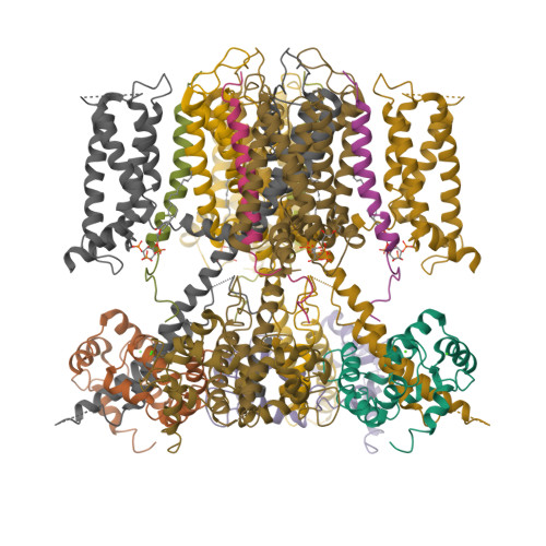
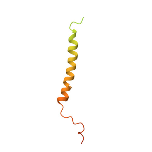
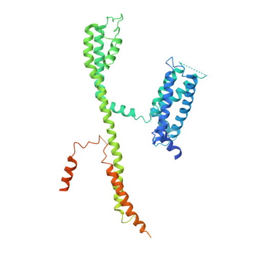
Structural Basis of Human KCNQ1 Modulation and Gating.

Sun, J.MacKinnon, R.

(2020) Cell 180: 340-347.e9

  • DOI: https://doi.org/10.1016/j.cell.2019.12.003
  • Primary Citation of Related Structures:  
    6UZZ, 6V00, 6V01

  • PubMed Abstract: 

    KCNQ1, also known as Kv7.1, is a voltage-dependent K + channel that regulates gastric acid secretion, salt and glucose homeostasis, and heart rhythm. Its functional properties are regulated in a tissue-specific manner through co-assembly with beta subunits KCNE1-5. In non-excitable cells, KCNQ1 forms a complex with KCNE3, which suppresses channel closure at negative membrane voltages that otherwise would close it. Pore opening is regulated by the signaling lipid PIP2. Using cryoelectron microscopy (cryo-EM), we show that KCNE3 tucks its single-membrane-spanning helix against KCNQ1, at a location that appears to lock the voltage sensor in its depolarized conformation. Without PIP2, the pore remains closed. Upon addition, PIP2 occupies a site on KCNQ1 within the inner membrane leaflet, which triggers a large conformational change that leads to dilation of the pore's gate. It is likely that this mechanism of PIP2 activation is conserved among Kv7 channels.


  • Organizational Affiliation

    Laboratory of Molecular Neurobiology and Biophysics and Howard Hughes Medical Institute, The Rockefeller University, 1230 York Avenue, New York, NY 10065, USA.


Macromolecules
Find similar proteins by:  (by identity cutoff)  |  3D Structure
Entity ID: 1
MoleculeChains Sequence LengthOrganismDetailsImage
Calmodulin-1A [auth B],
C [auth E],
E [auth H],
G [auth K]
149Homo sapiensMutation(s): 0 
Gene Names: CALM1CALMCAMCAM1
UniProt & NIH Common Fund Data Resources
Find proteins for P0DP23 (Homo sapiens)
Explore P0DP23 
Go to UniProtKB:  P0DP23
PHAROS:  P0DP23
GTEx:  ENSG00000198668 
Entity Groups  
Sequence Clusters30% Identity50% Identity70% Identity90% Identity95% Identity100% Identity
UniProt GroupP0DP23
Sequence Annotations
Expand
  • Reference Sequence
Find similar proteins by:  (by identity cutoff)  |  3D Structure
Entity ID: 2
MoleculeChains Sequence LengthOrganismDetailsImage
Potassium voltage-gated channel subfamily E member 3B [auth C],
D [auth F],
F [auth I],
H [auth L]
103Homo sapiensMutation(s): 0 
Gene Names: KCNE3
Membrane Entity: Yes 
UniProt & NIH Common Fund Data Resources
Find proteins for Q9Y6H6 (Homo sapiens)
Explore Q9Y6H6 
Go to UniProtKB:  Q9Y6H6
PHAROS:  Q9Y6H6
GTEx:  ENSG00000175538 
Entity Groups  
Sequence Clusters30% Identity50% Identity70% Identity90% Identity95% Identity100% Identity
UniProt GroupQ9Y6H6
Sequence Annotations
Expand
  • Reference Sequence
Find similar proteins by:  (by identity cutoff)  |  3D Structure
Entity ID: 3
MoleculeChains Sequence LengthOrganismDetailsImage
Potassium voltage-gated channel subfamily KQT member 1I [auth J],
J [auth A],
K [auth D],
L [auth G]
557Homo sapiensMutation(s): 0 
Gene Names: KCNQ1KCNA8KCNA9KVLQT1
Membrane Entity: Yes 
UniProt & NIH Common Fund Data Resources
Find proteins for P51787 (Homo sapiens)
Explore P51787 
Go to UniProtKB:  P51787
PHAROS:  P51787
GTEx:  ENSG00000053918 
Entity Groups  
Sequence Clusters30% Identity50% Identity70% Identity90% Identity95% Identity100% Identity
UniProt GroupP51787
Sequence Annotations
Expand
  • Reference Sequence
Small Molecules
Ligands 2 Unique
IDChains Name / Formula / InChI Key2D Diagram3D Interactions
PT5 (Subject of Investigation/LOI)
Query on PT5

Download Ideal Coordinates CCD File 
U [auth J],
V [auth A],
W [auth D],
X [auth G]
[(2R)-1-octadecanoyloxy-3-[oxidanyl-[(1R,2R,3S,4R,5R,6S)-2,3,6-tris(oxidanyl)-4,5-diphosphonooxy-cyclohexyl]oxy-phospho ryl]oxy-propan-2-yl] (8Z)-icosa-5,8,11,14-tetraenoate
C47 H85 O19 P3
CNWINRVXAYPOMW-HJBQCNPJSA-N
CA
Query on CA

Download Ideal Coordinates CCD File 
M [auth B]
N [auth B]
O [auth E]
P [auth E]
Q [auth H]
M [auth B],
N [auth B],
O [auth E],
P [auth E],
Q [auth H],
R [auth H],
S [auth K],
T [auth K]
CALCIUM ION
Ca
BHPQYMZQTOCNFJ-UHFFFAOYSA-N
Experimental Data & Validation

Experimental Data

  • Method: ELECTRON MICROSCOPY
  • Resolution: 3.90 Å
  • Aggregation State: PARTICLE 
  • Reconstruction Method: SINGLE PARTICLE 
EM Software:
TaskSoftware PackageVersion
RECONSTRUCTIONRELION

Structure Validation

View Full Validation Report


Entry History & Funding Information

Deposition Data


Funding OrganizationLocationGrant Number
National Institutes of Health/National Heart, Lung, and Blood InstituteUnited States5K99HL143037
Howard Hughes Medical InstituteUnited States--

Revision History  (Full details and data files)

  • Version 1.0: 2019-12-04
    Type: Initial release
  • Version 1.1: 2019-12-18
    Changes: Author supporting evidence
  • Version 1.2: 2020-01-15
    Changes: Database references
  • Version 1.3: 2020-02-05
    Changes: Database references
  • Version 2.0: 2024-03-20
    Changes: Advisory, Atomic model, Author supporting evidence, Data collection, Database references, Derived calculations, Polymer sequence, Source and taxonomy, Structure summary