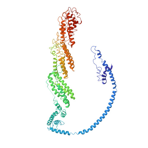
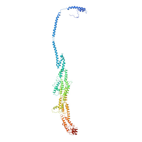
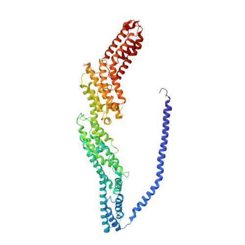
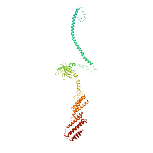
6VKL

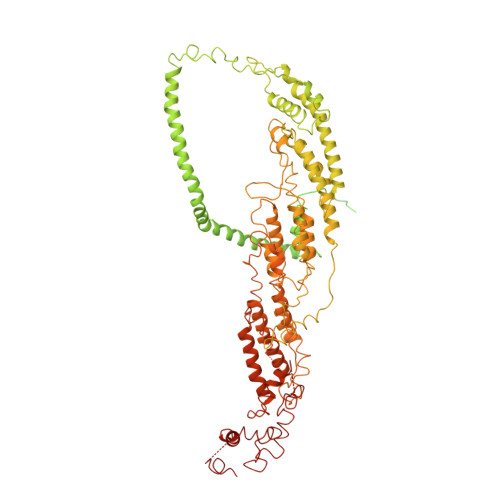
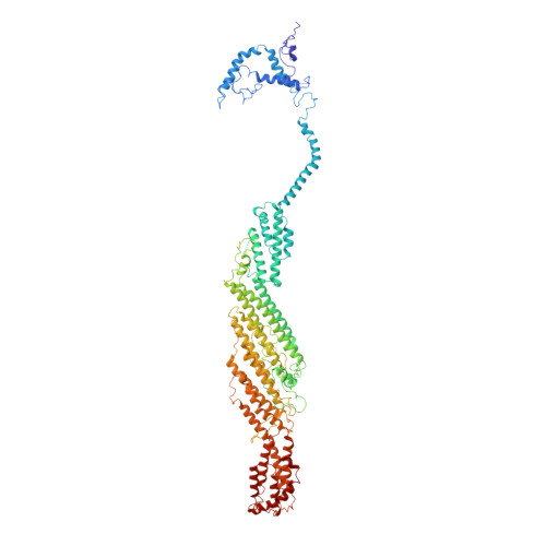
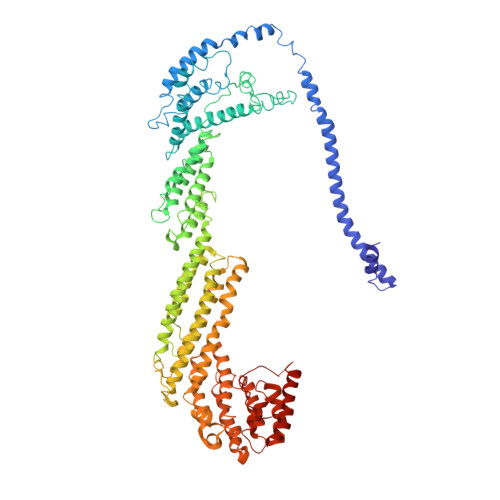
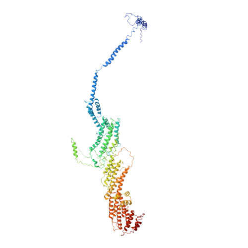
Negative stain reconstruction of the yeast exocyst octameric complex.


Experimental Data Snapshot

  • Method: ELECTRON MICROSCOPY
  • Resolution: 15.0 Å
  • Resolution: 15.0 Å
  • Aggregation State: PARTICLE 
  • Reconstruction Method: SINGLE PARTICLE 

wwPDB Validation   3D Report Full Report


This is version 1.1 of the entry. See complete history


Literature

Exocyst structural changes associated with activation of tethering downstream of Rho/Cdc42 GTPases.

Rossi, G.Lepore, D.Kenner, L.Czuchra, A.B.Plooster, M.Frost, A.Munson, M.Brennwald, P.

(2020) J Cell Biol 219

  • DOI: https://doi.org/10.1083/jcb.201904161
  • Primary Citation of Related Structures:  
    6VKL

  • PubMed Abstract: 

    The exocyst complex plays a critical role in determining both temporal and spatial dynamics of exocytic vesicle tethering and fusion with the plasma membrane. However, the mechanism by which the exocyst functions and how it is regulated remain poorly understood. Here we describe a novel biochemical assay for the examination of exocyst function in vesicle tethering. Importantly, the assay is stimulated by gain-of-function mutations in the Exo70 component of the exocyst, selected for their ability to bypass Rho/Cdc42 activation in vivo. Single-particle electron microscopy and 3D reconstructions of negatively stained exocyst complexes reveal a structural change in the mutant exocyst that exposes a binding site for the v-SNARE. We demonstrate a v-SNARE requirement in our tethering assay and increased v-SNARE binding to exocyst gain-of-function complexes. Together, these data suggest an allosteric mechanism for activation involving a conformational change in one subunit of the complex, which is relayed through the complex to regulate its biochemical activity in vitro, as well as overall function in vivo.


  • Organizational Affiliation

    Department of Cell Biology and Physiology, University of North Carolina School of Medicine, Chapel Hill, NC.


Macromolecules
Find similar proteins by:  (by identity cutoff)  |  3D Structure
Entity ID: 1
MoleculeChains Sequence LengthOrganismDetailsImage
Exocyst complex component SEC31,336Saccharomyces cerevisiae S288CMutation(s): 0 
UniProt
Find proteins for P33332 (Saccharomyces cerevisiae (strain ATCC 204508 / S288c))
Explore P33332 
Go to UniProtKB:  P33332
Entity Groups  
Sequence Clusters30% Identity50% Identity70% Identity90% Identity95% Identity100% Identity
UniProt GroupP33332
Sequence Annotations
Expand
  • Reference Sequence
Find similar proteins by:  (by identity cutoff)  |  3D Structure
Entity ID: 2
MoleculeChains Sequence LengthOrganismDetailsImage
Exocyst complex component SEC5971Saccharomyces cerevisiae S288CMutation(s): 0 
UniProt
Find proteins for P89102 (Saccharomyces cerevisiae (strain ATCC 204508 / S288c))
Explore P89102 
Go to UniProtKB:  P89102
Entity Groups  
Sequence Clusters30% Identity50% Identity70% Identity90% Identity95% Identity100% Identity
UniProt GroupP89102
Sequence Annotations
Expand
  • Reference Sequence
Find similar proteins by:  (by identity cutoff)  |  3D Structure
Entity ID: 3
MoleculeChains Sequence LengthOrganismDetailsImage
Exocyst complex component SEC6805Saccharomyces cerevisiae S288CMutation(s): 0 
UniProt
Find proteins for P32844 (Saccharomyces cerevisiae (strain ATCC 204508 / S288c))
Explore P32844 
Go to UniProtKB:  P32844
Entity Groups  
Sequence Clusters30% Identity50% Identity70% Identity90% Identity95% Identity100% Identity
UniProt GroupP32844
Sequence Annotations
Expand
  • Reference Sequence
Find similar proteins by:  (by identity cutoff)  |  3D Structure
Entity ID: 4
MoleculeChains Sequence LengthOrganismDetailsImage
Exocyst complex component SEC81,065Saccharomyces cerevisiae S288CMutation(s): 0 
UniProt
Find proteins for P32855 (Saccharomyces cerevisiae (strain ATCC 204508 / S288c))
Explore P32855 
Go to UniProtKB:  P32855
Entity Groups  
Sequence Clusters30% Identity50% Identity70% Identity90% Identity95% Identity100% Identity
UniProt GroupP32855
Sequence Annotations
Expand
  • Reference Sequence
Find similar proteins by:  (by identity cutoff)  |  3D Structure
Entity ID: 5
MoleculeChains Sequence LengthOrganismDetailsImage
Exocyst complex component SEC10871Saccharomyces cerevisiae S288CMutation(s): 0 
UniProt
Find proteins for Q06245 (Saccharomyces cerevisiae (strain ATCC 204508 / S288c))
Explore Q06245 
Go to UniProtKB:  Q06245
Entity Groups  
Sequence Clusters30% Identity50% Identity70% Identity90% Identity95% Identity100% Identity
UniProt GroupQ06245
Sequence Annotations
Expand
  • Reference Sequence
Find similar proteins by:  (by identity cutoff)  |  3D Structure
Entity ID: 6
MoleculeChains Sequence LengthOrganismDetailsImage
Exocyst complex component SEC15910Saccharomyces cerevisiae S288CMutation(s): 0 
UniProt
Find proteins for P22224 (Saccharomyces cerevisiae (strain ATCC 204508 / S288c))
Explore P22224 
Go to UniProtKB:  P22224
Entity Groups  
Sequence Clusters30% Identity50% Identity70% Identity90% Identity95% Identity100% Identity
UniProt GroupP22224
Sequence Annotations
Expand
  • Reference Sequence
Find similar proteins by:  (by identity cutoff)  |  3D Structure
Entity ID: 7
MoleculeChains Sequence LengthOrganismDetailsImage
Exocyst complex component EXO70623Saccharomyces cerevisiae S288CMutation(s): 0 
UniProt
Find proteins for P19658 (Saccharomyces cerevisiae (strain ATCC 204508 / S288c))
Explore P19658 
Go to UniProtKB:  P19658
Entity Groups  
Sequence Clusters30% Identity50% Identity70% Identity90% Identity95% Identity100% Identity
UniProt GroupP19658
Sequence Annotations
Expand
  • Reference Sequence
Find similar proteins by:  (by identity cutoff)  |  3D Structure
Entity ID: 8
MoleculeChains Sequence LengthOrganismDetailsImage
Exocyst complex component EXO84753Saccharomyces cerevisiae S288CMutation(s): 0 
UniProt
Find proteins for P38261 (Saccharomyces cerevisiae (strain ATCC 204508 / S288c))
Explore P38261 
Go to UniProtKB:  P38261
Entity Groups  
Sequence Clusters30% Identity50% Identity70% Identity90% Identity95% Identity100% Identity
UniProt GroupP38261
Sequence Annotations
Expand
  • Reference Sequence
Experimental Data & Validation

Experimental Data

  • Method: ELECTRON MICROSCOPY
  • Resolution: 15.0 Å
  • Resolution: 15.0 Å
  • Aggregation State: PARTICLE 
  • Reconstruction Method: SINGLE PARTICLE 

Structure Validation

View Full Validation Report


Entry History & Funding Information

Deposition Data


Funding OrganizationLocationGrant Number
National Institutes of Health/National Institute of General Medical Sciences (NIH/NIGMS)United States2R01GM068803

Revision History  (Full details and data files)

  • Version 1.0: 2020-07-29
    Type: Initial release
  • Version 1.1: 2024-03-06
    Changes: Author supporting evidence, Data collection, Database references, Refinement description