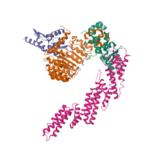
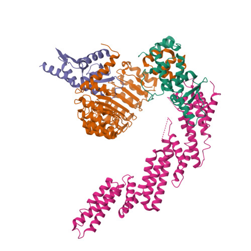
6WCQ

Structure of a substrate-bound DQC ubiquitin ligase


Experimental Data Snapshot

  • Method: ELECTRON MICROSCOPY
  • Resolution: 8.50 Å
  • Aggregation State: PARTICLE 
  • Reconstruction Method: SINGLE PARTICLE 

Starting Models: experimental
View more details

wwPDB Validation   3D Report Full Report


This is version 1.3 of the entry. See complete history


Literature

Structural basis for dimerization quality control.

Mena, E.L.Jevtic, P.Greber, B.J.Gee, C.L.Lew, B.G.Akopian, D.Nogales, E.Kuriyan, J.Rape, M.

(2020) Nature 586: 452-456

  • DOI: https://doi.org/10.1038/s41586-020-2636-7
  • Primary Citation of Related Structures:  
    6W66, 6W67, 6W68, 6W69, 6WCQ

  • PubMed Abstract: 

    Most quality control pathways target misfolded proteins to prevent toxic aggregation and neurodegeneration 1 . Dimerization quality control further improves proteostasis by eliminating complexes of aberrant composition 2 , but how it detects incorrect subunits remains unknown. Here we provide structural insight into target selection by SCF-FBXL17, a dimerization-quality-control E3 ligase that ubiquitylates and helps to degrade inactive heterodimers of BTB proteins while sparing functional homodimers. We find that SCF-FBXL17 disrupts aberrant BTB dimers that fail to stabilize an intermolecular β-sheet around a highly divergent β-strand of the BTB domain. Complex dissociation allows SCF-FBXL17 to wrap around a single BTB domain, resulting in robust ubiquitylation. SCF-FBXL17 therefore probes both shape and complementarity of BTB domains, a mechanism that is well suited to establish quality control of complex composition for recurrent interaction modules.


  • Organizational Affiliation

    Department of Molecular and Cell Biology, University of California at Berkeley, Berkeley, CA, USA.


Macromolecules
Find similar proteins by:  (by identity cutoff)  |  3D Structure
Entity ID: 1
MoleculeChains Sequence LengthOrganismDetailsImage
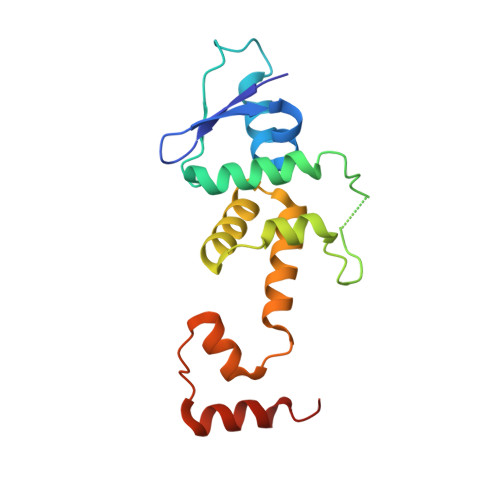
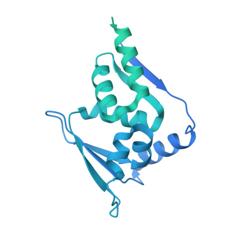
S-phase kinase-associated protein 1163Homo sapiensMutation(s): 0 
Gene Names: SKP1EMC19OCP2SKP1ATCEB1L
UniProt & NIH Common Fund Data Resources
Find proteins for P63208 (Homo sapiens)
Explore P63208 
Go to UniProtKB:  P63208
PHAROS:  P63208
GTEx:  ENSG00000113558 
Entity Groups  
Sequence Clusters30% Identity50% Identity70% Identity90% Identity95% Identity100% Identity
UniProt GroupP63208
Sequence Annotations
Expand
  • Reference Sequence
Find similar proteins by:  (by identity cutoff)  |  3D Structure
Entity ID: 2
MoleculeChains Sequence LengthOrganismDetailsImage
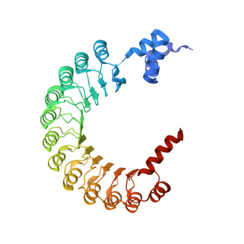
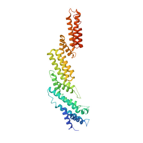
F-box/LRR-repeat protein 17396Homo sapiensMutation(s): 0 
Gene Names: FBXL17FBL17FBX13FBXO13
UniProt & NIH Common Fund Data Resources
Find proteins for Q9UF56 (Homo sapiens)
Explore Q9UF56 
Go to UniProtKB:  Q9UF56
PHAROS:  Q9UF56
GTEx:  ENSG00000145743 
Entity Groups  
Sequence Clusters30% Identity50% Identity70% Identity90% Identity95% Identity100% Identity
UniProt GroupQ9UF56
Sequence Annotations
Expand
  • Reference Sequence
Find similar proteins by:  (by identity cutoff)  |  3D Structure
Entity ID: 3
MoleculeChains Sequence LengthOrganismDetailsImage
Kelch-like ECH-associated protein 1631Homo sapiensMutation(s): 1 
Gene Names: KEAP1INRF2KIAA0132KLHL19
UniProt & NIH Common Fund Data Resources
Find proteins for Q14145 (Homo sapiens)
Explore Q14145 
Go to UniProtKB:  Q14145
PHAROS:  Q14145
GTEx:  ENSG00000079999 
Entity Groups  
Sequence Clusters30% Identity50% Identity70% Identity90% Identity95% Identity100% Identity
UniProt GroupQ14145
Sequence Annotations
Expand
  • Reference Sequence
Find similar proteins by:  (by identity cutoff)  |  3D Structure
Entity ID: 4
MoleculeChains Sequence LengthOrganismDetailsImage
Cullin-1434Homo sapiensMutation(s): 0 
Gene Names: CUL1
UniProt & NIH Common Fund Data Resources
Find proteins for Q13616 (Homo sapiens)
Explore Q13616 
Go to UniProtKB:  Q13616
PHAROS:  Q13616
GTEx:  ENSG00000055130 
Entity Groups  
Sequence Clusters30% Identity50% Identity70% Identity90% Identity95% Identity100% Identity
UniProt GroupQ13616
Sequence Annotations
Expand
  • Reference Sequence
Experimental Data & Validation

Experimental Data

  • Method: ELECTRON MICROSCOPY
  • Resolution: 8.50 Å
  • Aggregation State: PARTICLE 
  • Reconstruction Method: SINGLE PARTICLE 
EM Software:
TaskSoftware PackageVersion
RECONSTRUCTIONRELION3
MODEL REFINEMENTPHENIX

Structure Validation

View Full Validation Report


Entry History & Funding Information

Deposition Data


Funding OrganizationLocationGrant Number
Howard Hughes Medical Institute (HHMI)United States--

Revision History  (Full details and data files)

  • Version 1.0: 2020-08-19
    Type: Initial release
  • Version 1.1: 2020-09-02
    Changes: Database references
  • Version 1.2: 2020-10-28
    Changes: Database references
  • Version 1.3: 2024-03-06
    Changes: Data collection, Database references, Refinement description