6X9O

High resolution cryoEM structure of huntingtin in complex with HAP40


Experimental Data Snapshot

  • Method: ELECTRON MICROSCOPY
  • Resolution: 2.60 Å
  • Aggregation State: PARTICLE 
  • Reconstruction Method: SINGLE PARTICLE 

wwPDB Validation   3D Report Full Report


This is version 1.1 of the entry. See complete history


Literature

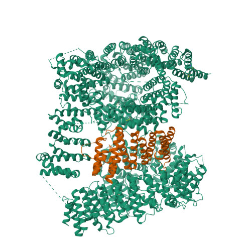
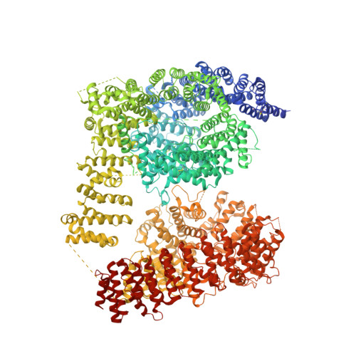
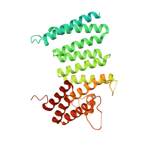
Huntingtin structure is orchestrated by HAP40 and shows a polyglutamine expansion-specific interaction with exon 1.

Harding, R.J.Deme, J.C.Hevler, J.F.Tamara, S.Lemak, A.Cantle, J.P.Szewczyk, M.M.Begeja, N.Goss, S.Zuo, X.Loppnau, P.Seitova, A.Hutchinson, A.Fan, L.Truant, R.Schapira, M.Carroll, J.B.Heck, A.J.R.Lea, S.M.Arrowsmith, C.H.

(2021) Commun Biol 4: 1374-1374

  • DOI: https://doi.org/10.1038/s42003-021-02895-4
  • Primary Citation of Related Structures:  
    6X9O

  • PubMed Abstract: 

    Huntington's disease results from expansion of a glutamine-coding CAG tract in the huntingtin (HTT) gene, producing an aberrantly functioning form of HTT. Both wildtype and disease-state HTT form a hetero-dimer with HAP40 of unknown functional relevance. We demonstrate in vivo and in cell models that HTT and HAP40 cellular abundance are coupled. Integrating data from a 2.6 Å cryo-electron microscopy structure, cross-linking mass spectrometry, small-angle X-ray scattering, and modeling, we provide a near-atomic-level view of HTT, its molecular interaction surfaces and compacted domain architecture, orchestrated by HAP40. Native mass spectrometry reveals a remarkably stable hetero-dimer, potentially explaining the cellular inter-dependence of HTT and HAP40. The exon 1 region of HTT is dynamic but shows greater conformational variety in the polyglutamine expanded mutant than wildtype exon 1. Our data provide a foundation for future functional and drug discovery studies targeting Huntington's disease and illuminate the structural consequences of HTT polyglutamine expansion.


  • Organizational Affiliation

    Structural Genomics Consortium, University of Toronto, Toronto, ON, M5G 1L7, Canada. Rachel.Harding@utoronto.ca.


Macromolecules
Find similar proteins by:  (by identity cutoff)  |  3D Structure
Entity ID: 1
MoleculeChains Sequence LengthOrganismDetailsImage
Huntingtin3,156Homo sapiensMutation(s): 2 
Gene Names: HTTHDIT15
UniProt & NIH Common Fund Data Resources
Find proteins for P42858 (Homo sapiens)
Explore P42858 
Go to UniProtKB:  P42858
PHAROS:  P42858
GTEx:  ENSG00000197386 
Entity Groups  
Sequence Clusters30% Identity50% Identity70% Identity90% Identity95% Identity100% Identity
UniProt GroupP42858
Sequence Annotations
Expand
  • Reference Sequence
Find similar proteins by:  (by identity cutoff)  |  3D Structure
Entity ID: 2
MoleculeChains Sequence LengthOrganismDetailsImage
40-kDa huntingtin-associated protein389Homo sapiensMutation(s): 0 
Gene Names: F8A1F8A2F8A3
UniProt & NIH Common Fund Data Resources
Find proteins for P23610 (Homo sapiens)
Explore P23610 
Go to UniProtKB:  P23610
GTEx:  ENSG00000277150 
Entity Groups  
Sequence Clusters30% Identity50% Identity70% Identity90% Identity95% Identity100% Identity
UniProt GroupP23610
Sequence Annotations
Expand
  • Reference Sequence
Experimental Data & Validation

Experimental Data

  • Method: ELECTRON MICROSCOPY
  • Resolution: 2.60 Å
  • Aggregation State: PARTICLE 
  • Reconstruction Method: SINGLE PARTICLE 
EM Software:
TaskSoftware PackageVersion
RECONSTRUCTIONRELION3.1

Structure Validation

View Full Validation Report


Entry History 

Deposition Data

Revision History  (Full details and data files)

  • Version 1.0: 2020-06-17
    Type: Initial release
  • Version 1.1: 2022-03-30
    Changes: Database references