6Y3C

Human COX-1 Crystal Structure


Experimental Data Snapshot

  • Method: X-RAY DIFFRACTION
  • Resolution: 3.36 Å
  • R-Value Free: 0.263 
  • R-Value Work: 0.208 
  • R-Value Observed: 0.214 

wwPDB Validation   3D Report Full Report


This is version 2.2 of the entry. See complete history


Literature

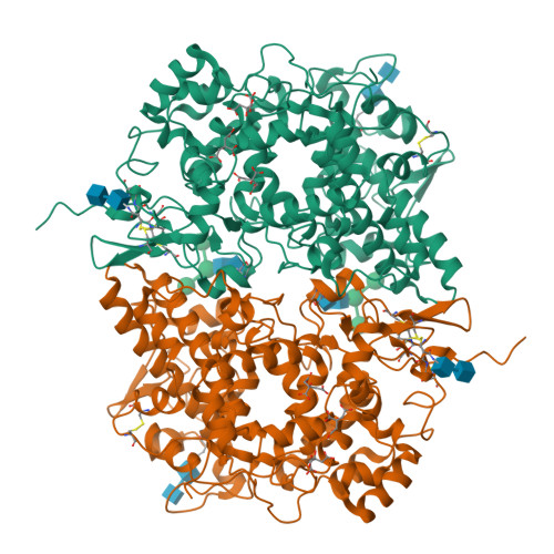
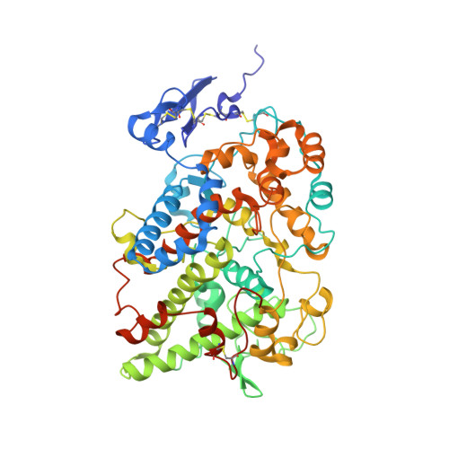
Three-dimensional structure of human cyclooxygenase (hCOX)-1.

Miciaccia, M.Belviso, B.D.Iaselli, M.Cingolani, G.Ferorelli, S.Cappellari, M.Loguercio Polosa, P.Perrone, M.G.Caliandro, R.Scilimati, A.

(2021) Sci Rep 11: 4312-4312

  • DOI: https://doi.org/10.1038/s41598-021-83438-z
  • Primary Citation of Related Structures:  
    6Y3C

  • PubMed Abstract: 

    The beneficial effects of Cyclooxygenases (COX) inhibitors on human health have been known for thousands of years. Nevertheless, COXs, particularly COX-1, have been linked to a plethora of human diseases such as cancer, heart failure, neurological and neurodegenerative diseases only recently. COXs catalyze the first step in the biosynthesis of prostaglandins (PGs) and are among the most important mediators of inflammation. All published structural work on COX-1 deals with the ovine isoenzyme, which is easier to produce in milligram-quantities than the human enzyme and crystallizes readily. Here, we report the long-sought structure of the human cyclooxygenase-1 (hCOX-1) that we refined to an R/R free of 20.82/26.37, at 3.36 Å resolution. hCOX-1 structure provides a detailed picture of the enzyme active site and the residues crucial for inhibitor/substrate binding and catalytic activity. We compared hCOX-1 crystal structure with the ovine COX-1 and human COX-2 structures by using metrics based on Cartesian coordinates, backbone dihedral angles, and solvent accessibility coupled with multivariate methods. Differences and similarities among structures are discussed, with emphasis on the motifs responsible for the diversification of the various enzymes (primary structure, stability, catalytic activity, and specificity). The structure of hCOX-1 represents an essential step towards the development of new and more selective COX-1 inhibitors of enhanced therapeutic potential.


  • Organizational Affiliation

    Department of Pharmacy - Pharmaceutical Sciences, University of Bari "Aldo Moro", Via E. Orabona 4, 70125, Bari, Italy.


Macromolecules
Find similar proteins by:  (by identity cutoff)  |  3D Structure
Entity ID: 1
MoleculeChains Sequence LengthOrganismDetailsImage
Prostaglandin G/H synthase 1576Homo sapiensMutation(s): 0 
Gene Names: PTGS1COX1
EC: 1.14.99.1
Membrane Entity: Yes 
UniProt & NIH Common Fund Data Resources
Find proteins for P23219 (Homo sapiens)
Explore P23219 
Go to UniProtKB:  P23219
PHAROS:  P23219
GTEx:  ENSG00000095303 
Entity Groups  
Sequence Clusters30% Identity50% Identity70% Identity90% Identity95% Identity100% Identity
UniProt GroupP23219
Sequence Annotations
Expand
  • Reference Sequence
Oligosaccharides

Help

Entity ID: 2
MoleculeChains Length2D Diagram Glycosylation3D Interactions
beta-D-mannopyranose-(1-3)-[beta-D-mannopyranose-(1-6)]beta-D-mannopyranose-(1-4)-2-acetamido-2-deoxy-beta-D-glucopyranose-(1-4)-2-acetamido-2-deoxy-beta-D-glucopyranose
B
5N-Glycosylation
Glycosylation Resources
GlyTouCan:  G89225LT
GlyCosmos:  G89225LT
GlyGen:  G89225LT
Entity ID: 3
MoleculeChains Length2D Diagram Glycosylation3D Interactions
2-acetamido-2-deoxy-beta-D-glucopyranose-(1-4)-2-acetamido-2-deoxy-beta-D-glucopyranose
C, D
2N-Glycosylation
Glycosylation Resources
GlyTouCan:  G42666HT
GlyCosmos:  G42666HT
GlyGen:  G42666HT
Experimental Data & Validation

Experimental Data

  • Method: X-RAY DIFFRACTION
  • Resolution: 3.36 Å
  • R-Value Free: 0.263 
  • R-Value Work: 0.208 
  • R-Value Observed: 0.214 
  • Space Group: P 65 2 2
Unit Cell:
Length ( Å )Angle ( ˚ )
a = 182.11α = 90
b = 182.11β = 90
c = 103.24γ = 120
Software Package:
Software NamePurpose
SCALAdata scaling
PHENIXrefinement
PDB_EXTRACTdata extraction
xia2data reduction
Sir2014phasing

Structure Validation

View Full Validation Report


Entry History 

Deposition Data

Revision History  (Full details and data files)

  • Version 1.0: 2020-02-26
    Type: Initial release
  • Version 1.1: 2020-03-11
    Changes: Database references
  • Version 1.2: 2020-04-08
    Changes: Refinement description
  • Version 2.0: 2020-07-29
    Type: Remediation
    Reason: Carbohydrate remediation
    Changes: Advisory, Atomic model, Data collection, Derived calculations, Structure summary
  • Version 2.1: 2021-03-03
    Changes: Database references, Structure summary
  • Version 2.2: 2024-01-24
    Changes: Data collection, Database references, Refinement description