7JW7

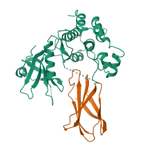
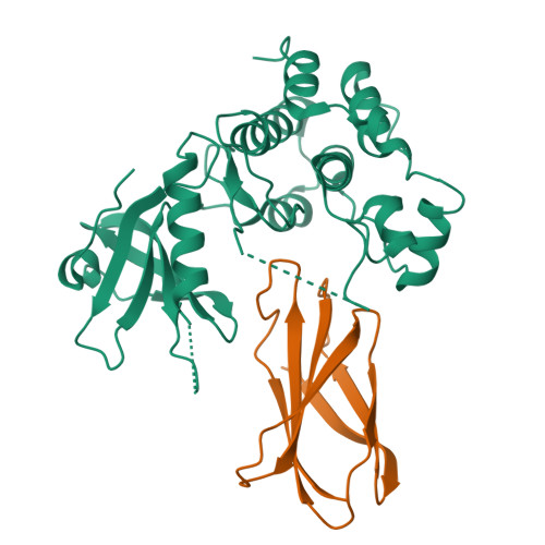
Structure of monobody 27 human MLKL pseudokinase domain complex


Experimental Data Snapshot

  • Method: X-RAY DIFFRACTION
  • Resolution: 2.63 Å
  • R-Value Free: 0.281 
  • R-Value Work: 0.224 
  • R-Value Observed: 0.230 

Starting Models: experimental
View more details

wwPDB Validation   3D Report Full Report


This is version 1.2 of the entry. See complete history


Literature

Conformational interconversion of MLKL and disengagement from RIPK3 precede cell death by necroptosis.

Garnish, S.E.Meng, Y.Koide, A.Sandow, J.J.Denbaum, E.Jacobsen, A.V.Yeung, W.Samson, A.L.Horne, C.R.Fitzgibbon, C.Young, S.N.Smith, P.P.C.Webb, A.I.Petrie, E.J.Hildebrand, J.M.Kannan, N.Czabotar, P.E.Koide, S.Murphy, J.M.

(2021) Nat Commun 12: 2211-2211

  • DOI: https://doi.org/10.1038/s41467-021-22400-z
  • Primary Citation of Related Structures:  
    7JW7, 7JXU

  • PubMed Abstract: 

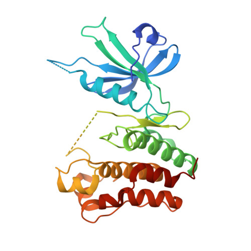
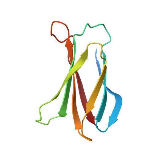
    Phosphorylation of the MLKL pseudokinase by the RIPK3 kinase leads to MLKL oligomerization, translocation to, and permeabilization of, the plasma membrane to induce necroptotic cell death. The precise choreography of MLKL activation remains incompletely understood. Here, we report Monobodies, synthetic binding proteins, that bind the pseudokinase domain of MLKL within human cells and their crystal structures in complex with the human MLKL pseudokinase domain. While Monobody-32 constitutively binds the MLKL hinge region, Monobody-27 binds MLKL via an epitope that overlaps the RIPK3 binding site and is only exposed after phosphorylated MLKL disengages from RIPK3 following necroptotic stimulation. The crystal structures identified two distinct conformations of the MLKL pseudokinase domain, supporting the idea that a conformational transition accompanies MLKL disengagement from RIPK3. These studies provide further evidence that MLKL undergoes a large conformational change upon activation, and identify MLKL disengagement from RIPK3 as a key regulatory step in the necroptosis pathway.


  • Organizational Affiliation

    Walter and Eliza Hall Institute of Medical Research, Parkville, VIC, Australia.


Macromolecules
Find similar proteins by:  (by identity cutoff)  |  3D Structure
Entity ID: 1
MoleculeChains Sequence LengthOrganismDetailsImage
Mixed lineage kinase domain-like protein287Homo sapiensMutation(s): 0 
Gene Names: MLKL
UniProt & NIH Common Fund Data Resources
Find proteins for Q8NB16 (Homo sapiens)
Explore Q8NB16 
Go to UniProtKB:  Q8NB16
PHAROS:  Q8NB16
GTEx:  ENSG00000168404 
Entity Groups  
Sequence Clusters30% Identity50% Identity70% Identity90% Identity95% Identity100% Identity
UniProt GroupQ8NB16
Sequence Annotations
Expand
  • Reference Sequence
Find similar proteins by:  (by identity cutoff)  |  3D Structure
Entity ID: 2
MoleculeChains Sequence LengthOrganismDetailsImage
Monobody 2798synthetic constructMutation(s): 0 
Entity Groups  
Sequence Clusters30% Identity50% Identity70% Identity90% Identity95% Identity100% Identity
Sequence Annotations
Expand
  • Reference Sequence
Experimental Data & Validation

Experimental Data

  • Method: X-RAY DIFFRACTION
  • Resolution: 2.63 Å
  • R-Value Free: 0.281 
  • R-Value Work: 0.224 
  • R-Value Observed: 0.230 
  • Space Group: P 31 2 1
Unit Cell:
Length ( Å )Angle ( ˚ )
a = 64.805α = 90
b = 64.805β = 90
c = 226.763γ = 120
Software Package:
Software NamePurpose
PHENIXrefinement
Aimlessdata scaling
PDB_EXTRACTdata extraction
XDSdata reduction
PHENIXphasing

Structure Validation

View Full Validation Report


Entry History & Funding Information

Deposition Data


Funding OrganizationLocationGrant Number
National Health and Medical Research Council (NHMRC, Australia)Australia1172929
National Health and Medical Research Council (NHMRC, Australia)Australia1124735

Revision History  (Full details and data files)

  • Version 1.0: 2021-03-10
    Type: Initial release
  • Version 1.1: 2021-04-28
    Changes: Database references
  • Version 1.2: 2023-10-18
    Changes: Data collection, Database references, Refinement description