
1SNS (replaced by 2SNS):
A High Resolution Structure of an Inhibitor Complex of the Extracellular Nuclease of Staphylococcus aureus: I. EXPERIMENTAL PROCEDURES AND CHAIN TRACING.
A. Arnone, C.J. Bier, F.A. Cotton, V.W. Day, E.E. Hazen Jr., D.C. Richardson, J.S. Richardson, A. Yonath 1971
|

2cna:
The covalent and three-dimensional structure of concanavalin A. IV. Atomic coordinates, hydrogen bonding, and quaternary structure.
G N Reeke Jr, J W Becker, G M Edelman 1975
|

1gpd:
Studies of asymmetry in the three-dimensional structure of lobster D-glyceraldehyde-3-phosphate dehydrogenase.
D Moras, KW Olsen, MN Sabesan, M Buehner, GC Ford, MG Rossmann 1975
|

4DFR:
Crystal structures of Escherichia coli and Lactobacillus casei dihydrofolate reductase refined at 1.7 A resolution. I. General features and binding of methotrexate.
J T Bolin, D J Filman, D A Matthews, R C Hamlin, J Kraut 1982
|

3BNA:
Reversible bending and helix geometry in a B-DNA dodecamer: CGCGAATTBrCGCG.
A V Fratini, M L Kopka, H R Drew, R E Dickerson 1982
|

2CYP:
Crystal structure of yeast cytochrome c peroxidase refined at 1.7-A resolution.
B C Finzel, T L Poulos, J Kraut 1984
|
1HBS:
Refined crystal structure of deoxyhemoglobin S. I. Restrained least-squares refinement at 3.0-A resolution.
DE A Padlan, W E Love 1985
|

1PP2:
The refined crystal structure of dimeric phospholipase A2 at 2.5 A. Access to a shielded catalytic center.
S Brunie, J Bolin, D Gewirth, P B Sigler 1985
|

3ICB:
The refined structure of vitamin D-dependent calcium-binding protein from bovine intestine. Molecular details, ion binding, and implications for the structure of other calcium-binding proteins.
D M Szebenyi, K Moffat 1986
|

5ADH:
Interdomain motion in liver alcohol dehydrogenase. Structural and energetic analysis of the hinge bending mode.
F Colonna-Cesari, D Perahia, M Karplus, H Eklund, C I Brädén, O Tapia 1986
|

113D:
The structure of guanosine-thymidine mismatches in B-DNA at 2.5-A resolution.
W.N. Hunter, T. Brown, G. Kneale, N.N. Anand, D. Rabinovich, O. Kennard 1987
|

4TNC:
Refined structure of chicken skeletal muscle troponin C in the two-calcium state at 2-A resolution.
K A Satyshur, S T Rao, D Pyzalska, W Drendel, M Greaser, M Sundaralingam 1988
|

1DCG:
The molecular structure of the left-handed Z-DNA double helix at 1.0-Å atomic resolution: Geometry, conformation, and ionic interactions of d(CGCGCG).
R V Gessner, C A Frederick, G J Quigley, A Rich, A H J Wang 1989
|

1TNF:
The Structure of Tumor Necrosis Factor-α at 2.6 Å Resolution: Implications for receptor binding.
M J Eck, S R Sprang 1989
|

2GLS:
Refined atomic model of glutamine synthetase at 3.5 Å resolution.
M M Yamashita, R J Almassy, C A Janson, D Cascio, D Eisenberg 1989
|

5HVP:
Crystallographic analysis of a complex between human immunodeficiency virus type 1 protease and acetyl-pepstatin at 2.0-A resolution.
P.M. Fitzgerald, B.M. McKeever, J.F. VanMiddlesworth, J.P. Springer, J.C. Heimbach, C.T. Leu, W.K. Herber, R.A. Dixon, P.L. Darke 1990
|
1HEM:
Structural and thermodynamic analysis of compensating mutations within the core of chicken egg white lysozyme.
K.P. Wilson, B.A. Malcolm, B.W. Matthews 1992
|

1WAT:
The three-dimensional structure of the ligand-binding domain of a wild-type bacterial chemotaxis receptor. Structural comparison to the cross-linked mutant forms and conformational changes upon ligand binding.
J.I. Yeh, H.P. Biemann, J. Pandit, D.E. Koshland, S.H. Kim 1993
|
1M2C:
A New α-Conotoxin Which Targets α3β2 Nicotinic Acetylcholine Receptors.
G. Edward Cartier, Doju Yoshikami, William R. Gray, Siqin Luo, Baldomero M. Olivera, J. Michael McIntosh Eisenberg 1996
|

1EEM:
Identification, Characterization, and Crystal Structure of the Omega Class Glutathione Transferases.
Philip G. Board, Marjorie Coggan, Gareth Chelvanayagam, Simon Easteal, Lars S. Jermiin, Gayle K. Schulte, Dennis E. Danley, Lise R. Hoth, Matthew C. Griffor, Ajith V. Kamath, Michele H. Rosner, Boris A. Chrunyk, David E. Perregaux, Christopher A. Gabel, Kieran F. Geoghegan, Jayvardhan Pandit 2000
|

1E3K, 1E3G:
Structural Evidence for Ligand Specificity in the Binding Domain of the Human Androgen Receptor: IMPLICATIONS FOR PATHOGENIC GENE MUTATIONS.
Pedro M. Matias, Peter Donner, Ricardo Coelho, Monica Thomaz, Cristina Peixoto, Sofia Macedo, Norbert Otto, Simone Joschko, Peter Scholz, Anja Wegg, Siegfried Bäsler, Martina Schäfer, Ursula Egner, Maria Arménia Carrondo 2000
|

1E78, 1E7B, 1E7A, 1E7C:
Binding of the General Anesthetics Propofol and Halothane to Human Serum Albumin: HIGH RESOLUTION CRYSTAL STRUCTURES.
Ananyo A. Bhattacharya, Stephen Curry, Nicholas P. Franks 2000
|

1DXR:
Structural Basis of the Drastically Increased Initial Electron Transfer Rate in the Reaction Center from a Rhodopseudomonas viridis Mutant Described at 2.00-Å Resolution.
C. Roy D. Lancaster, Marina V. Bibikova, Piera Sabatino, Dieter Oesterhelt, Hartmut Michel 2000
|

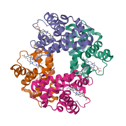
1FX0:
The Structure of the Chloroplast F1-ATPase at 3.2 Å Resolution.
Georg Groth, Ehmke Pohl 2001
|

1HA2, 1H9Z:
Crystal Structure Analysis of Warfarin Binding to Human Serum Albumin: ANATOMY OF DRUG SITE I.
Isabelle Petitpas, Ananyo A. Bhattacharya, Sue Twine, Malcolm East, Stephen Curry 2001
|

1HUY:
Reducing the Environmental Sensitivity of Yellow Fluorescent Protein: MECHANISM AND APPLICATIONS.
Oliver Griesbeck, Geoffrey S. Baird, Robert E. Campbell, David A. Zacharias, Roger Y. Tsien 2001
|

1L0V:
Crystallographic Studies of the Escherichia coli Quinol-Fumarate Reductase with Inhibitors Bound to the Quinol-binding Site.
Tina M. Iverson, César Luna-Chavez, Laura R. Croal, Gary Cecchini, Douglas C. Rees 2002
|

1GYC:
Crystal Structure of a Laccase from the FungusTrametes versicolor at 1.90-Å Resolution Containing a Full Complement of Coppers.
Klaus Piontek, Matteo Antorini, Thomas Choinowski 2002
|

1M14, 1M17:
Structure of the Epidermal Growth Factor Receptor Kinase Domain Alone and in Complex with a 4-Anilinoquinazoline Inhibitor.
Jennifer Stamos, Mark X. Sliwkowski, Charles Eigenbrot 2002
|

1P0P, 1P0M, 1P0I, 1P0Q:
Crystal Structure of Human Butyrylcholinesterase and of Its Complexes with Substrate and Products.
Yvain Nicolet, Oksana Lockridge, Patrick Masson, Juan C. Fontecilla-Camps, Florian Nachon 2003
|

1Q5K:
Structural Insights and Biological Effects of Glycogen Synthase Kinase 3-specific Inhibitor AR-A014418.
Ratan Bhat, Yafeng Xue, Stefan Berg, Sven Hellberg, Mats Ormö, Yvonne Nilsson, Ann-Cathrin Radesäter, Eva Jerning, Per-Olof Markgren, Thomas Borgegård, Martin Nylöf, Alfredo Giménez-Cassina, Félix Hernández, Jose J. Lucas, Javier Díaz-Nido, Jesús Avila 2003
|

1JC1, 1JC0:
Investigating Mitochondrial Redox Potential with Redox-sensitive Green Fluorescent Protein Indicators.
George T. Hanson, Robert Aggeler, Devin Oglesbee, Mark Cannon, Roderick A. Capaldi, Roger Y. Tsien, S. James Remington 2004
|

1VF7:
Crystal Structure of the Membrane Fusion Protein, MexA, of the Multidrug Transporter in Pseudomonas aeruginosa.
Hiroyuki Akama, Takanori Matsuura, Sachiko Kashiwagi, Hiroshi Yoneyama, Shin-ichiro Narita, Tomitake Tsukihara, Atsushi Nakagawa, Taiji Nakae 2004
|

1T45, 1T46:
Structural Basis for the Autoinhibition and STI-571 Inhibition of c-Kit Tyrosine Kinase.
Clifford D. Mol, Douglas R. Dougan, Thomas R. Schneider, Robert J. Skene, Michelle L. Kraus, Daniel N. Scheibe, Gyorgy P. Snell, Hua Zou, Bi-Ching Sang, Keith P. Wilson 2004
|

1R9O:
The Structure of Human Cytochrome P450 2C9 Complexed with Flurbiprofen at 2.0-Å Resolution.
Michael R. Wester, Jason K. Yano, Guillaume A. Schoch, Christine Yang, Keith J. Griffin, C. David Stout, Eric F. Johnson 2004
|

1TQN:
The Structure of Human Microsomal Cytochrome P450 3A4 Determined by X-ray Crystallography to 2.05-Å Resolution.
Jason K. Yano, Michael R. Wester, Guillaume A. Schoch, Keith J. Griffin, C. David Stout, Eric F. Johnson 2004
|

1R7G, 1R7F, 1R7E, 1R7D, 1R7C:
Structure and Function of the Membrane Anchor Domain of Hepatitis C Virus Nonstructural Protein 5A.
François Penin, Volker Brass, Nicole Appel, Stephanie Ramboarina, Roland Montserret, Damien Ficheux, Hubert E. Blum, Ralf Bartenschlager, Darius Moradpour 2004
|

1XQ8:
Structure and Dynamics of Micelle-bound Human α-Synuclein.
Tobias S. Ulmer, Ad Bax, Nelson B. Cole, Robert L. Nussbaum, Published in issue: March 11, 2005
|

2ZMX, 2AHL, 2AHK, 1WXC, 1WX5, 1WX4, 1WX2:
Crystallographic Evidence That the Dinuclear Copper Center of Tyrosinase Is Flexible during Catalysis.
Yasuyuki Matoba, Takanori Kumagai, Aiko Yamamoto, Hironari Yoshitsu, Masanori Sugiyama 2006
|

2P1L:
Crystal Structure of the Bcl-XL-Beclin 1 Peptide Complex: BECLIN 1 IS A NOVEL BH3-ONLY PROTEIN.
Adam Oberstein, Philip D. Jeffrey, Yigong Shi 2007
|

2ZJD:
Structural Basis for Sorting Mechanism of p62 in Selective Autophagy.
Yoshinobu Ichimura, Taichi Kumanomidou, Yu-shin Sou, Tsunehiro Mizushima, Junji Ezaki, Takashi Ueno, Eiki Kominami, Takashi Yamane, Keiji Tanaka, Masaaki Komatsu 2008
|
|
|
|


