


Full Text |
QUERY: PDB Deposit Group ID(s) = G_1002115 | MyPDB Login | Search API |
| Search Summary | This query matches 7 Structures. |
Structure Determination MethodologyScientific Name of Source OrganismTaxonomyExperimental MethodPolymer Entity TypeRefinement Resolution (Å)Release DateEnzyme Classification NameSymmetry TypeSCOP Classification | 1 to 7 of 7 Structures Page 1 of 1 Sort by 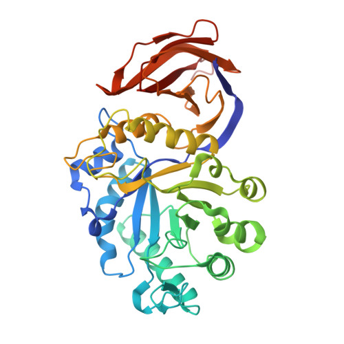
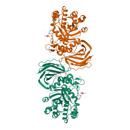
Crystal Structure of XynC from Bacillus subtilis 168St John, F.J., Hurlbert, J.C., Pozharski, E. (2009) Acta Crystallogr Sect F Struct Biol Cryst Commun 65: 499-503
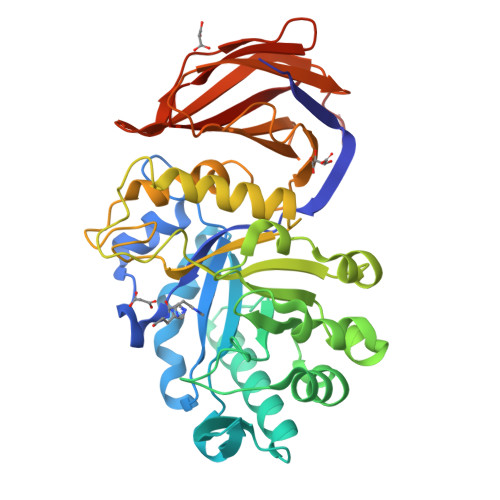
Crystal structure of the glucuronoxylan xylanohydrolase XynC from Bacillus subtilisSt John, F.J., Hurlbert, J.C., Pozharski, E. (2011) J Mol Biology 407: 92-109

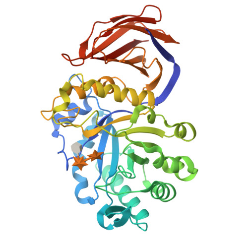
Crystal structure of Ligand bound XynCSt John, F.J., Hurlbert, J.C., Pozharski, E. (2011) J Mol Biology 407: 92-109
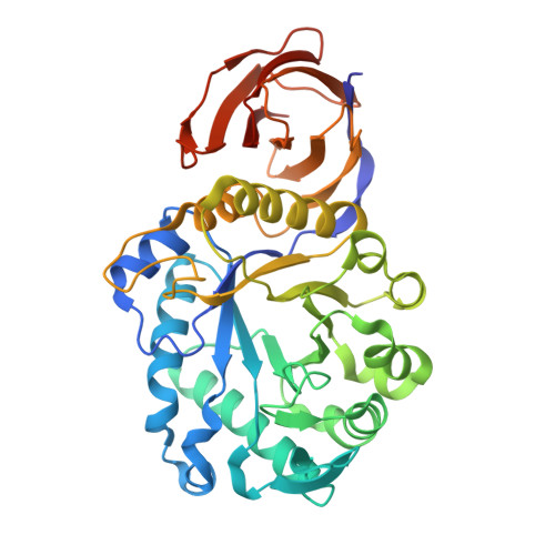
Structure Analysis of a Xylanase From Glycosyl Hydrolase Family Thirty: Carbohydrate Ligand Complexes Reveal this Family of Enzymes Unique Mechanism of Substrate Specificity and RecognitionSt John, F.J., Hurlbert, J.C., Pozharski, E. (2011) J Mol Biology 407: 92-109
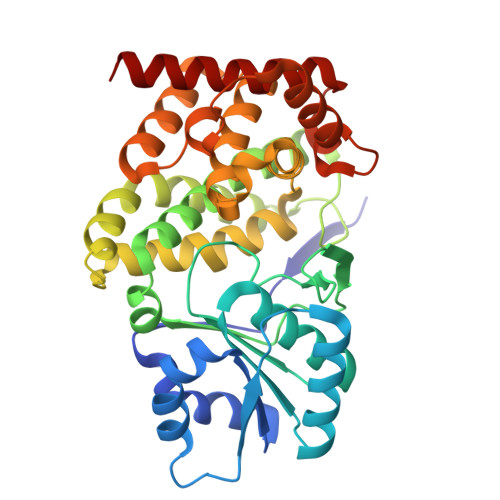
Crystal Structure Analysis of a GH30 Endoxylanase from Clostridium papyrosolvens C71Bales, E.B., Smith, J.K., St John, F.J., Hurlbert, J.C. (2014) Acta Crystallogr D Biol Crystallogr 70: 2950-2958
X-ray crystallographic protein structure of the glycoside hydrolase family 30 subfamily 8 xylanase, Xyn30A, from Clostridium acetobutylicumSt John, F.J., Pozharski, E., Hurlbert, J.C. To be published
Structure of a Bacillus coagulans polyol dehydrogenase double mutant with an acquired D-lactate dehydrogenase activity(2020) Protein Sci 29: 2387-2397
1 to 7 of 7 Structures Page 1 of 1 Sort by |

