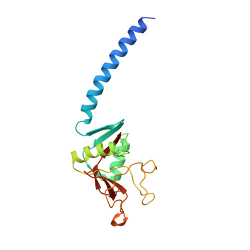
1B08

LUNG SURFACTANT PROTEIN D (SP-D) (FRAGMENT)


Experimental Data Snapshot

  • Method: X-RAY DIFFRACTION
  • Resolution: 2.30 Å
  • R-Value Free: 0.271 
  • R-Value Work: 0.209 
  • R-Value Observed: 0.209 

Starting Model: experimental
View more details

wwPDB Validation   3D Report Full Report


This is version 1.4 of the entry. See complete history


Literature

Crystal structure of the trimeric alpha-helical coiled-coil and the three lectin domains of human lung surfactant protein D.

Hakansson, K.Lim, N.K.Hoppe, H.J.Reid, K.B.

(1999) Structure 7: 255-264

  • DOI: https://doi.org/10.1016/s0969-2126(99)80036-7
  • Primary Citation of Related Structures:  
    1B08

  • PubMed Abstract: 

    Human lung surfactant protein D (hSP-D) belongs to the collectin family of C-type lectins and participates in the innate immune surveillance against microorganisms in the lung through recognition of carbohydrate ligands present on the surface of pathogens. The involvement of this protein in innate immunity and the allergic response make it the subject of much interest. We have determined the crystal structure of a trimeric fragment of hSP-D at 2.3 A resolution. The structure comprises an alpha-helical coiled-coil and three carbohydrate-recognition domains (CRDs). An interesting deviation from symmetry was found in the projection of a single tyrosine sidechain into the centre of the coiled-coil; the asymmetry of this residue influences the orientation of one of the adjacent CRDs. The cleft between the three CRDs presents a large positively charged surface. The fold of the CRD of hSP-D is similar to that of the mannan-binding protein (MBP), but its orientation relative to the alpha-helical coiled-coil region differs somewhat to that seen in the MBP structure. The novel central packing of the tyrosine sidechain within the coiled-coil and the resulting asymmetric orientation of the CRDs has unexpected functional implications. The positively charged surface might facilitate binding to negatively charged structures, such as lipopolysaccharides.


  • Organizational Affiliation

    Department of Microbiology, University of Illinois at Urbana-Champaign, B103 CLSL, 601 South Goodwin Avenue, Urbana, IL 61801, USA. kjell@scs.uiuc.edu


Macromolecules
Find similar proteins by:  (by identity cutoff)  |  3D Structure
Entity ID: 1
MoleculeChains Sequence LengthOrganismDetailsImage
PROTEIN (LUNG SURFACTANT PROTEIN D)
A, B, C
158Homo sapiensMutation(s): 0 
UniProt & NIH Common Fund Data Resources
Find proteins for P35247 (Homo sapiens)
Explore P35247 
Go to UniProtKB:  P35247
PHAROS:  P35247
GTEx:  ENSG00000133661 
Entity Groups  
Sequence Clusters30% Identity50% Identity70% Identity90% Identity95% Identity100% Identity
UniProt GroupP35247
Sequence Annotations
Expand
  • Reference Sequence
Small Molecules
Ligands 1 Unique
IDChains Name / Formula / InChI Key2D Diagram3D Interactions
CA
Query on CA

Download Ideal Coordinates CCD File 
D [auth A]
E [auth A]
F [auth A]
G [auth B]
H [auth B]
D [auth A],
E [auth A],
F [auth A],
G [auth B],
H [auth B],
I [auth B],
J [auth C],
K [auth C],
L [auth C]
CALCIUM ION
Ca
BHPQYMZQTOCNFJ-UHFFFAOYSA-N
Experimental Data & Validation

Experimental Data

  • Method: X-RAY DIFFRACTION
  • Resolution: 2.30 Å
  • R-Value Free: 0.271 
  • R-Value Work: 0.209 
  • R-Value Observed: 0.209 
  • Space Group: P 1 21 1
Unit Cell:
Length ( Å )Angle ( ˚ )
a = 55.96α = 90
b = 109.72β = 92.2
c = 56.09γ = 90
Software Package:
Software NamePurpose
DENZOdata reduction
SCALEPACKdata scaling
CCP4model building
X-PLORrefinement
AMoREphasing

Structure Validation

View Full Validation Report


Entry History 

Deposition Data

Revision History  (Full details and data files)

  • Version 1.0: 1999-12-01
    Type: Initial release
  • Version 1.1: 2007-10-16
    Changes: Version format compliance
  • Version 1.2: 2011-07-13
    Changes: Derived calculations, Version format compliance
  • Version 1.3: 2023-08-09
    Changes: Data collection, Database references, Derived calculations, Refinement description
  • Version 1.4: 2024-11-06
    Changes: Structure summary