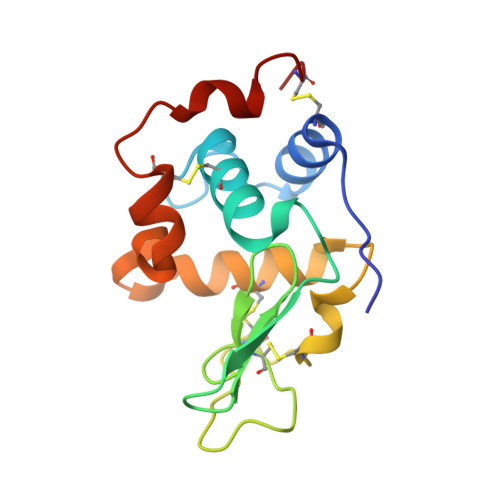
Effect of extra N-terminal residues on the stability and folding of human lysozyme expressed in Pichia pastoris.
Goda, S., Takano, K., Yamagata, Y., Katakura, Y., Yutani, K.(2000) Protein Eng 13: 299-307
- PubMed: 10810162
- DOI: https://doi.org/10.1093/protein/13.4.299
- Primary Citation of Related Structures:
1C7P - PubMed Abstract:
A human lysozyme expression system by Pichia pastoris was constructed with the expression vector of pPIC9, which contains the alpha-factor signal peptide known for high secretion efficiency. P. pastoris expressed the human lysozyme at about 300 mg/l broth, but four extra residues (Glu(-4)-Ala(-3)-Glu(-2)-Ala(-1)-) were added at the N-terminus of the expressed protein (EAEA-lysozyme). To determine the effect of the four extra residues on the stability, structures and folding of the protein, calorimetry, X-ray crystal analysis and GuHCl denaturation experiments were performed. The calorimetric studies showed that the EAEA-lysozyme was destabilized by 9.6 kJ/mol at pH 2.7 compared with the wild-type protein, mainly caused by the substantial decrease in the enthalpy change (DeltaH). On the basis of structural information on the EAEA-lysozyme, thermodynamic analyses show that (1) the addition of the four residues slightly affected the conformation in other parts far from the N-terminus, (2) the large decrease in the enthalpy change due to the conformational changes would be almost compensated by the decrease in the entropy change and (3) the decrease in the Gibbs energy change between the EAEA and wild-type human lysozymes could be explained by the summation of each Gibbs energy change contributing to the stabilizing factors concerning the extra residues.
- Institute for Protein Research, Osaka University, Yamadaoka, Suita, Osaka 565-0871, Japan.
Organizational Affiliation:


