1C7U | pdb_00001c7u

Complex of the DNA binding core domain of the transcription factor MEF2A with a 20mer oligonucleotide


Experimental Data Snapshot

  • Method: SOLUTION NMR
  • Conformers Calculated: 35 
  • Conformers Submitted: 
  • Selection Criteria: REGULARIZED MEAN STRUCTURE 

wwPDB Validation 3D Report Full Report

Validation slider image for 1C7U

This is version 1.4 of the entry. See complete history

Literature

Solution structure of the MEF2A-DNA complex: structural basis for the modulation of DNA bending and specificity by MADS-box transcription factors

Huang, K.Louis, J.M.Donaldson, L.Lim, F.L.Sharrocks, A.D.Clore, G.M.

(2000) EMBO J 19: 2615-2628

  • DOI: https://doi.org/10.1093/emboj/19.11.2615
  • Primary Citation Related Structures: 
    1C7U

  • PubMed Abstract: 

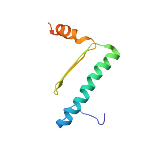
    The solution structure of the 33 kDa complex between the dimeric DNA-binding core domain of the transcription factor MEF2A (residues 1-85) and a 20mer DNA oligonucleotide comprising the consensus sequence CTA(A/T)(4)TAG has been solved by NMR. The protein comprises two domains: a MADS-box (residues 1-58) and a MEF2S domain (residues 59-73). Recognition and specificity are achieved by interactions between the MADS-box and both the major and minor grooves of the DNA. A number of critical differences in protein-DNA contacts observed in the MEF2A-DNA complex and the DNA complexes of the related MADS-box transcription factors SRF and MCM1 provide a molecular explanation for modulation of sequence specificity and extent of DNA bending ( approximately 15 versus approximately 70 degrees ). The structure of the MEF2S domain is entirely different from that of the equivalent SAM domain in SRF and MCM1, accounting for the absence of cross-reactivity with other proteins that interact with these transcription factors.


  • Organizational Affiliation
    • Laboratory of Chemical Physics, Building 5, National Institute of Diabetes and Digestive and Kidney Diseases, National Institutes of Health, Bethesda, MD 20892-0510, USA.

Macromolecule Content 

  • Total Structure Weight: 32.28 kDa 
  • Atom Count: 2,038 
  • Modeled Residue Count: 188 
  • Deposited Residue Count: 210 
  • Unique protein chains: 1
  • Unique nucleic acid chains: 1

Macromolecules


Find similar proteins by:|  3D Structure
Entity ID: 2
MoleculeChains  Sequence LengthOrganismDetailsImage
MYOCYTE-SPECIFIC ENHANCER FACTOR 2A, C4 FORMC [auth A],
D [auth B]
85Homo sapiensMutation(s): 0 
UniProt & NIH Common Fund Data Resources
Find proteins for Q02078 (Homo sapiens)
Explore Q02078 
Go to UniProtKB:  Q02078
PHAROS:  Q02078
GTEx:  ENSG00000068305 
Entity Groups
Sequence Clusters30% Identity50% Identity70% Identity90% Identity95% Identity100% Identity
UniProt GroupQ02078
Sequence Annotations
Expand
Reference Sequence
Find similar nucleic acids by:  Sequence
Entity ID: 1
MoleculeChains LengthOrganismImage
5'-D(*CP*TP*CP*GP*GP*CP*TP*AP*TP*TP*AP*AP*TP*AP*GP*CP*CP*GP*AP*G)-3'A [auth C],
B [auth D]
20N/A
Sequence Annotations
Expand
Reference Sequence

Experimental Data & Validation

Experimental Data

  • Method: SOLUTION NMR
  • Conformers Calculated: 35 
  • Conformers Submitted: 
  • Selection Criteria: REGULARIZED MEAN STRUCTURE 

Structure Validation

View Full Validation Report


Entry History 

Deposition Data

Revision History  (Full details and data files)

  • Version 1.0: 2000-03-27
    Type: Initial release
  • Version 1.1: 2008-04-26
    Changes: Version format compliance
  • Version 1.2: 2011-07-13
    Changes: Version format compliance
  • Version 1.3: 2022-02-16
    Changes: Database references, Derived calculations
  • Version 1.4: 2023-12-27
    Changes: Data collection