1CQJ

CRYSTAL STRUCTURE OF DEPHOSPHORYLATED E. COLI SUCCINYL-COA SYNTHETASE


Experimental Data Snapshot

  • Method: X-RAY DIFFRACTION
  • Resolution: 2.90 Å
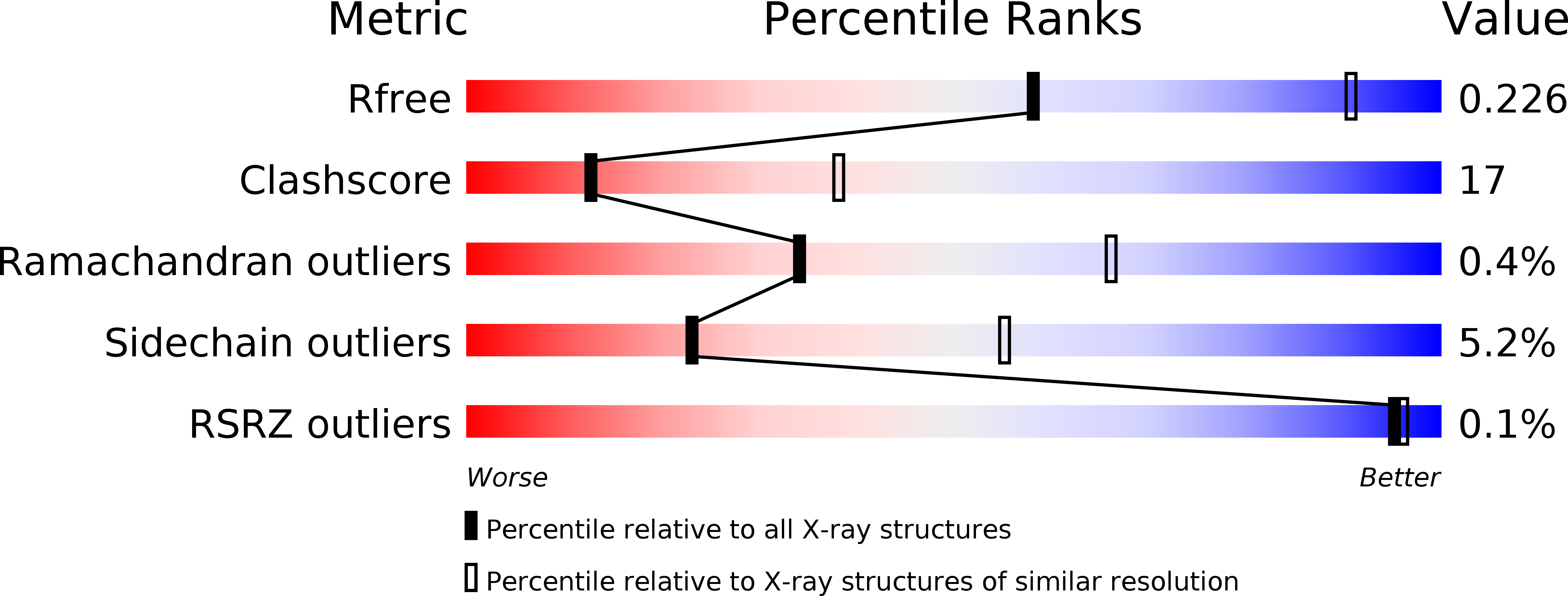
  • R-Value Free: 0.228 
  • R-Value Work: 0.172 
  • R-Value Observed: 0.178 

wwPDB Validation   3D Report Full Report


Ligand Structure Quality Assessment 


This is version 1.4 of the entry. See complete history


Literature

ADP-binding site of Escherichia coli succinyl-CoA synthetase revealed by x-ray crystallography.

Joyce, M.A.Fraser, M.E.James, M.N.Bridger, W.A.Wolodko, W.T.

(2000) Biochemistry 39: 17-25

  • DOI: https://doi.org/10.1021/bi991696f
  • Primary Citation of Related Structures:  
    1CQI, 1CQJ

  • PubMed Abstract: 

    Succinyl-CoA synthetase (SCS) catalyzes the following reversible reaction via a phosphorylated histidine intermediate (His 246alpha): succinyl-CoA + P(i) + NDP <--> succinate + CoA + NTP (N denotes adenosine or guanosine). To determine the structure of the enzyme with nucleotide bound, crystals of phosphorylated Escherichia coli SCS were soaked in successive experiments adopting progressive strategies. In the first experiment, 1 mM ADP (>15 x K(d)) was added; Mg(2+) ions were omitted to preclude the formation of an insoluble precipitate with the phosphate and ammonium ions. X-ray crystallography revealed that the enzyme was dephosphorylated, but the nucleotide did not remain bound to the enzyme (R(working) = 17.2%, R(free) = 22.8% for data to 2.9 A resolution). Catalysis requires Mg(2+) ions; hence, the "true" nucleotide substrate is probably an ADP-Mg(2+) complex. In the successful experiment, the phosphate buffer was exchanged with MOPS, the concentration of sulfate ions was lowered, and the concentrations of ADP and Mg(2+) ions were increased to 10.5 and 50 mM, respectively. X-ray diffraction data revealed an ADP-Mg(2+) complex bound in the ATP-grasp fold of the N-terminal domain of each beta-subunit (R(working) = 19.1%, R(free) = 24.7% for data to 3.3 A resolution). We describe the specific interactions of the nucleotide-Mg(2+) complex with SCS, compare these results with those for other proteins containing the ATP-grasp fold, and present a hypothetical model of the histidine-containing loop in the "down" position where it can interact with the nucleotide approximately 35 A from where His 246alpha is seen in both phosphorylated and dephosphorylated SCS.


  • Organizational Affiliation

    Department of Biochemistry, University of Alberta, Edmonton, Canada.


Macromolecules
Find similar proteins by:  (by identity cutoff)  |  3D Structure
Entity ID: 1
MoleculeChains Sequence LengthOrganismDetailsImage
SUCCINYL-COA SYNTHETASE ALPHA CHAINA,
C [auth D]
286Escherichia coliMutation(s): 0 
EC: 6.2.1.5
UniProt
Find proteins for P0AGE9 (Escherichia coli (strain K12))
Explore P0AGE9 
Go to UniProtKB:  P0AGE9
Entity Groups  
Sequence Clusters30% Identity50% Identity70% Identity90% Identity95% Identity100% Identity
UniProt GroupP0AGE9
Sequence Annotations
Expand
  • Reference Sequence
Find similar proteins by:  (by identity cutoff)  |  3D Structure
Entity ID: 2
MoleculeChains Sequence LengthOrganismDetailsImage
SUCCINYL-COA SYNTHETASE BETA CHAINB,
D [auth E]
385Escherichia coliMutation(s): 0 
EC: 6.2.1.5
UniProt
Find proteins for P0A836 (Escherichia coli (strain K12))
Explore P0A836 
Go to UniProtKB:  P0A836
Entity Groups  
Sequence Clusters30% Identity50% Identity70% Identity90% Identity95% Identity100% Identity
UniProt GroupP0A836
Sequence Annotations
Expand
  • Reference Sequence
Experimental Data & Validation

Experimental Data

  • Method: X-RAY DIFFRACTION
  • Resolution: 2.90 Å
  • R-Value Free: 0.228 
  • R-Value Work: 0.172 
  • R-Value Observed: 0.178 
  • Space Group: P 43 2 2
Unit Cell:
Length ( Å )Angle ( ˚ )
a = 98.82α = 90
b = 98.82β = 90
c = 404.68γ = 90
Software Package:
Software NamePurpose
BIOMOLmodel building
CNSrefinement
WEISdata reduction
SCALKB2data scaling
KBAPLYdata scaling

Structure Validation

View Full Validation Report


Ligand Structure Quality Assessment 


Entry History 

Deposition Data

Revision History  (Full details and data files)

  • Version 1.0: 2000-01-10
    Type: Initial release
  • Version 1.1: 2008-04-27
    Changes: Version format compliance
  • Version 1.2: 2011-07-13
    Changes: Version format compliance
  • Version 1.3: 2017-10-04
    Changes: Refinement description
  • Version 1.4: 2024-10-30
    Changes: Data collection, Database references, Derived calculations, Structure summary