1DB5 | pdb_00001db5

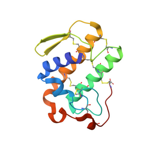
HUMAN S-PLA2 IN COMPLEX WITH INDOLE 6


Experimental Data Snapshot

  • Method: X-RAY DIFFRACTION
  • Resolution: 2.80 Å
  • R-Value Free: 
    0.240 (Depositor) 
  • R-Value Work: 
    0.196 (Depositor) 
  • R-Value Observed: 
    0.196 (Depositor) 

wwPDB Validation 3D Report Full Report

Validation slider image for 1DB5

This is version 1.4 of the entry. See complete history. 

Literature

Structure-based design of the first potent and selective inhibitor of human non-pancreatic secretory phospholipase A2.

Schevitz, R.W., Bach, N.J., Carlson, D.G., Chirgadze, N.Y., Clawson, D.K., Dillard, R.D., Draheim, S.E., Hartley, L.W., Jones, N.D., Mihelich, E.D., Olkowski, J.L., Snyder, D.W., Sommers, C., Wery, J.-P.

(1995) Nat Struct Biol 2: 458-465

  • DOI: https://doi.org/10.1038/nsb0695-458
  • Primary Citation Related Structures: 
    1DB4, 1DB5, 1DCY

  • PubMed Abstract: 

    A lead compound obtained from a high volume human non-pancreatic secretory phospholipase A2 (hnps-PLA2) screen has been developed into a potent inhibitor using detailed structural knowledge of inhibitor binding to the enzyme active site. Four crystal structures of hnps-PLA2 complexed with a series of increasingly potent indole inhibitors were determined and used as the structural basis for both understanding this binding and providing valuable insights for further development. The application of structure-based drug design has made possible improvements in the binding of this screening lead to the enzyme by nearly three orders of magnitude. Furthermore, the optimized structure (LY311727) displayed 1,500-fold selectivity when assayed against porcine pancreatic s-PLA2.


  • Organizational Affiliation: 
    • Lilly Research Laboratories, Lilly Corporate Center, Eli Lilly and Company, Indianapolis, Indiana 46285, USA.

Macromolecule Content 

  • Total Structure Weight: 14.41 kDa 
  • Atom Count: 1,034 
  • Modeled Residue Count: 124 
  • Deposited Residue Count: 124 
  • Unique protein chains: 1

Macromolecules

Find similar proteins by:|  3D Structure
Entity ID: 1
MoleculeChains  Sequence LengthOrganismDetailsImage
PROTEIN (PHOSPHOLIPASE A2)124Homo sapiensMutation(s): 0 
EC: 3.1.1.4
UniProt & NIH Common Fund Data Resources
Find proteins for P14555 (Homo sapiens)
Explore P14555 
Go to UniProtKB:  P14555
PHAROS:  P14555
GTEx:  ENSG00000188257 
Entity Groups
Sequence Clusters30% Identity50% Identity70% Identity90% Identity95% Identity100% Identity
UniProt GroupP14555
Sequence Annotations
Expand
Reference Sequence

Small Molecules

Ligands 2 Unique
IDChains Name / Formula / InChI Key2D Diagram3D Interactions
6IN

Query on 6IN


Download:Ideal Coordinates CCD File
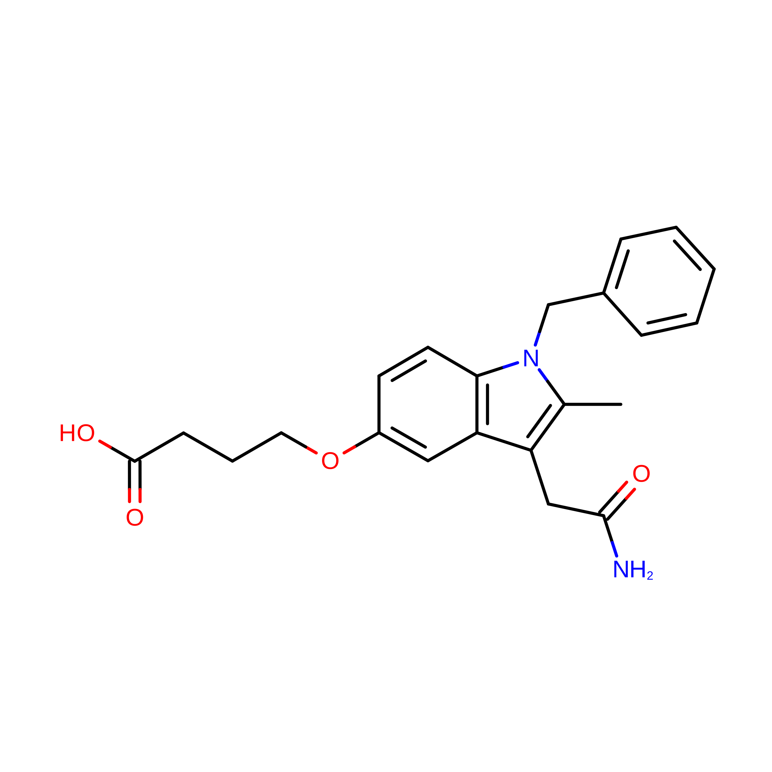
D [auth A]4-(1-BENZYL-3-CARBAMOYLMETHYL-2-METHYL-1H-INDOL-5-YLOXY)-BUTYRIC ACID
C22 H24 N2 O4
STENXUCYNKOBHJ-UHFFFAOYSA-N
CA

Query on CA


Download:Ideal Coordinates CCD File
B [auth A],
C [auth A]
CALCIUM ION
Ca
BHPQYMZQTOCNFJ-UHFFFAOYSA-N
Binding Affinity Annotations 
IDSourceBinding Affinity
6IN BindingDB:  1DB5 IC50: min: 152, max: 6.90e+4 (nM) from 3 assay(s)

Experimental Data & Validation

Experimental Data

  • Method: X-RAY DIFFRACTION
  • Resolution: 2.80 Å
  • R-Value Free:  0.240 (Depositor) 
  • R-Value Work:  0.196 (Depositor) 
  • R-Value Observed: 0.196 (Depositor) 
Space Group: P 61 2 2
Unit Cell:
Length ( Å )Angle ( ˚ )
a = 76.36α = 90
b = 76.36β = 90
c = 91.53γ = 120
Software Package:
Software NamePurpose
DENZOdata reduction
SCALEPACKdata scaling
X-PLORmodel building
X-PLORrefinement
X-PLORphasing

Structure Validation

View Full Validation Report


Entry History 

Deposition Data

Revision History  (Full details and data files)

  • Version 1.0: 1999-11-12
    Type: Initial release
  • Version 1.1: 2008-04-27
    Changes: Version format compliance
  • Version 1.2: 2011-07-13
    Changes: Version format compliance
  • Version 1.3: 2012-02-29
    Changes: Database references
  • Version 1.4: 2024-10-30
    Changes: Data collection, Database references, Derived calculations, Structure summary