1DCA | pdb_00001dca

STRUCTURE OF AN ENGINEERED METAL BINDING SITE IN HUMAN CARBONIC ANHYDRASE II REVEALS THE ARCHITECTURE OF A REGULATORY CYSTEINE SWITCH


Experimental Data Snapshot

  • Method: X-RAY DIFFRACTION
  • Resolution: 2.20 Å
  • R-Value Observed: 
    0.182 (Depositor) 

wwPDB Validation 3D Report Full Report

Validation slider image for 1DCA

This is version 1.4 of the entry. See complete history

Literature

Structure of an engineered His3Cys zinc binding site in human carbonic anhydrase II.

Ippolito, J.A.Christianson, D.W.

(1993) Biochemistry 32: 9901-9905

  • DOI: https://doi.org/10.1021/bi00089a005
  • Primary Citation Related Structures: 
    1DCA, 1DCB

  • PubMed Abstract: 

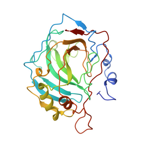
    X-ray crystallographic analysis of the Thr-199-->Cys (T199C) variant of human carbonic anhydrase II reveals the first high-resolution structure of an engineered zinc coordination polyhedron in a metalloenzyme. In the wild-type enzyme, Thr-199 accepts a hydrogen bond from zinc-bound hydroxide; in the variant, the polypeptide backbone is sufficiently plastic to permit Cys-199 to displace hydroxide ion and coordinate to zinc with nearly perfect coordination stereochemistry. Importantly, the resulting His3-Cys-Zn2+ motif binds zinc more tightly than the wild-type enzyme [Kiefer, L. L., Krebs, J. F., Paterno, S. A., & Fierke C. A. (1993) Biochemistry (preceding paper in this issue)]. This novel zinc coordination polyhedron is analogous to that postulated for matrix metalloproteinase zymogens such as prostromelysin, where a cysteine-zinc interaction is responsible for the inactivity of the zymogen. Intriguingly, Cys-199 of T199C CAII is displaced from zinc coordination by soaking crystals in high concentrations of acetazolamide. Hence, residual catalytic activity measured for this variant probably arises from an alternate conformer of Cys-199 which allows the catalytic nucleophile, hydroxide ion, to be activated by zinc coordination.


  • Organizational Affiliation
    • Department of Chemistry, University of Pennsylvania, Philadelphia 19104-6323.

Macromolecule Content 

  • Total Structure Weight: 29.36 kDa 
  • Atom Count: 2,129 
  • Modeled Residue Count: 255 
  • Deposited Residue Count: 260 
  • Unique protein chains: 1

Macromolecules

Find similar proteins by:|  3D Structure
Entity ID: 1
MoleculeChains  Sequence LengthOrganismDetailsImage
CARBONIC ANHYDRASE II260Homo sapiensMutation(s): 0 
EC: 4.2.1.1 (PDB Primary Data), 4.2.1.69 (UniProt)
UniProt & NIH Common Fund Data Resources
Find proteins for P00918 (Homo sapiens)
Explore P00918 
Go to UniProtKB:  P00918
PHAROS:  P00918
GTEx:  ENSG00000104267 
Entity Groups
Sequence Clusters30% Identity50% Identity70% Identity90% Identity95% Identity100% Identity
UniProt GroupP00918
Sequence Annotations
Expand
Reference Sequence

Small Molecules

Ligands 1 Unique
IDChains Name / Formula / InChI Key2D Diagram3D Interactions
ZN

Query on ZN


Download:Ideal Coordinates CCD File
B [auth A]ZINC ION
Zn
PTFCDOFLOPIGGS-UHFFFAOYSA-N

Experimental Data & Validation

Experimental Data

  • Method: X-RAY DIFFRACTION
  • Resolution: 2.20 Å
  • R-Value Observed: 0.182 (Depositor) 
Space Group: P 1 21 1
Unit Cell:
Length ( Å )Angle ( ˚ )
a = 42.7α = 90
b = 41.7β = 104.6
c = 73γ = 90
Software Package:
Software NamePurpose
PROLSQrefinement

Structure Validation

View Full Validation Report


Entry History 

Deposition Data

Revision History  (Full details and data files)

  • Version 1.0: 1993-07-15
    Type: Initial release
  • Version 1.1: 2008-03-24
    Changes: Version format compliance
  • Version 1.2: 2011-07-13
    Changes: Version format compliance
  • Version 1.3: 2017-11-29
    Changes: Derived calculations, Other
  • Version 1.4: 2024-02-07
    Changes: Data collection, Database references, Derived calculations