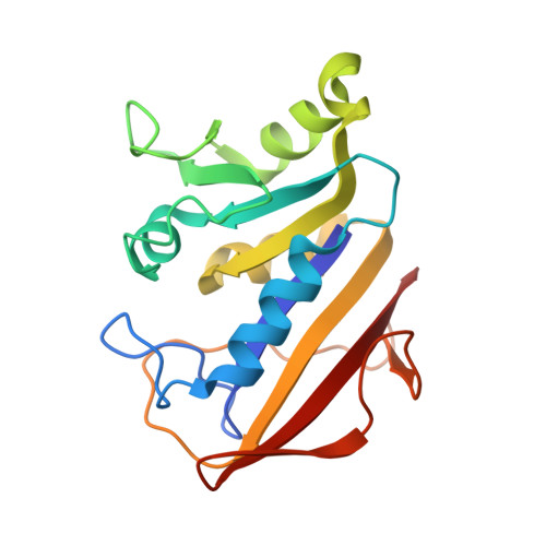
Crystal structures of recombinant human dihydrofolate reductase complexed with folate and 5-deazafolate.
Davies 2nd., J.F., Delcamp, T.J., Prendergast, N.J., Ashford, V.A., Freisheim, J.H., Kraut, J.(1990) Biochemistry 29: 9467-9479
- PubMed: 2248959
- DOI: https://doi.org/10.1021/bi00492a021
- Primary Citation of Related Structures:
1DHF, 2DHF - PubMed Abstract:
The 2.3-A crystal structure of recombinant human dihydrofolate reductase (EC 1.5.1.3, DHFR) has been solved as a binary complex with folate (a poor substrate at neutral pH) and also as a binary complex with an inhibitor, 5-deazafolate. The inhibitor appears to be protonated at N8 on binding, whereas folate is not. Rotation of the peptide plane joining I7 and V8 from its position in the folate complex permits hydrogen bonding of 5-deazafolate's protonated N8 to the backbone carbonyl of I7, thus contributing to the enzyme's greater affinity for 5-deazafolate than for folate. In this respect it is likely that bound 5-deazafolate furnishes a model for 7,8-dihydrofolate binding and, in addition, resembles the transition state for folate reduction. A hypothetical transition-state model for folate reduction, generated by superposition of the DHFR binary complexes human.5-deazafolate and chicken liver.NADPH, reveals a 1-A overlap of the binding sites for folate's pteridine ring and the dihydronicotinamide ring of NADPH. It is proposed that this binding-site overlap accelerates the reduction of both folate and 7,8-dihydrofolate by simultaneously binding substrate and cofactor with a sub van der Waals separation that is optimal for hydride transfer.
- Department of Chemistry, University of California, San Diego, La Jolla 92093.
Organizational Affiliation:


