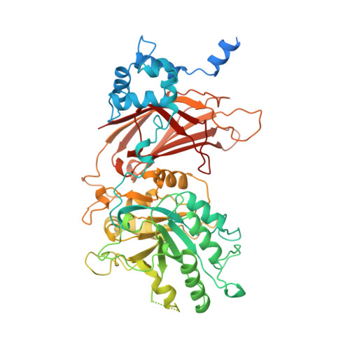
A ternary metal binding site in the C2 domain of phosphoinositide-specific phospholipase C-delta1.
Essen, L.O., Perisic, O., Lynch, D.E., Katan, M., Williams, R.L.(1997) Biochemistry 36: 2753-2762
- PubMed: 9062102
- DOI: https://doi.org/10.1021/bi962466t
- Primary Citation of Related Structures:
1DJG, 1DJH, 1DJI - PubMed Abstract:
We have determined the crystal structures of complexes of phosphoinositide-specific phospholipase C-delta1 from rat with calcium, barium, and lanthanum at 2.5-2.6 A resolution. Binding of these metal ions is observed in the active site of the catalytic TIM barrel and in the calcium binding region (CBR) of the C2 domain. The C2 domain of PLC-delta1 is a circularly permuted topological variant (P-variant) of the synaptotagmin I C2A domain (S-variant). On the basis of sequence analysis, we propose that both the S-variant and P-variant topologies are present among other C2 domains. Multiple adjacent binding sites in the C2 domain were observed for calcium and the other metal/enzyme complexes. The maximum number of binding sites observed was for the calcium analogue lanthanum. This complex shows an array-like binding of three lanthanum ions (sites I-III) in a crevice on one end of the C2 beta-sandwich. Residues involved in metal binding are contained in three loops, CBR1, CBR2, and CBR3. Sites I and II are maintained in the calcium and barium complexes, whereas sites II and III coincide with a binary calcium binding site in the C2A domain of synaptotagmin I. Several conformers for CBR1 are observed. The conformation of CBR1 does not appear to be strictly dependent on metal binding; however, metal binding may stabilize certain conformers. No significant structural changes are observed for CBR2 or CBR3. The surface of this ternary binding site provides a cluster of freely accessible liganding positions for putative phospholipid ligands of the C2 domain. It may be that the ternary metal binding site is also a feature of calcium-dependent phospholipid binding in solution. A ternary metal binding site might be a conserved feature among C2 domains that contain the critical calcium ligands in their CBR's. The high cooperativity of calcium-mediated lipid binding by C2 domains described previously is explained by this novel type of calcium binding site.
- Centre for Protein Engineering, MRC Centre, Cambridge, U.K.
Organizational Affiliation:


