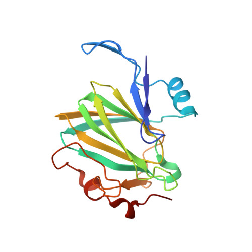
1DZT

RMLC FROM SALMONELLA TYPHIMURIUM


Experimental Data Snapshot

  • Method: X-RAY DIFFRACTION
  • Resolution: 2.20 Å
  • R-Value Free: 0.275 
  • R-Value Work: 0.232 
  • R-Value Observed: 0.232 

Starting Model: experimental
View more details

wwPDB Validation   3D Report Full Report


Ligand Structure Quality Assessment 


This is version 1.2 of the entry. See complete history


Literature

Rmlc, the Third Enzyme of Dtdp-L-Rhamnose Pathway, is a New Class of Epimerase.

Giraud, M.F.Leonard, G.A.Field, R.A.Berlind, C.Naismith, J.H.

(2000) Nat Struct Biol 7: 398

  • DOI: https://doi.org/10.1038/75178
  • Primary Citation of Related Structures:  
    1DZR, 1DZT

  • PubMed Abstract: 

    Deoxythymidine diphosphate (dTDP)-L-rhamnose is the precursor of L-rhamnose, a saccharide required for the virulence of some pathogenic bacteria. dTDP-L-rhamnose is synthesized from glucose-1-phosphate and deoxythymidine triphosphate (dTTP) via a pathway involving four distinct enzymes. This pathway does not exist in humans and the enzymes involved in dTDP-L-rhamnose synthesis are potential targets for the design of new therapeutic agents. Here, the crystal structure of dTDP-6-deoxy-D-xylo-4-hexulose 3,5 epimerase (RmlC, EC5.1.3.13) from Salmonella enterica serovar Typhimurium was determined. The third enzyme of the rhamnose biosynthetic pathway, RmlC epimerizes at two carbon centers, the 3 and 5 positions of the sugar ring. The structure was determined by multiwavelength anomalous diffraction to a resolution of 2.17 A. RmlC is a dimer and each monomer is formed mainly from two beta-sheets arranged in a beta-sandwich. The structure of a dTDP-phenol-RmlC complex shows the substrate-binding site to be located between the two beta-sheets; this site is formed from residues of both monomers. Sequence alignments of other RmlC enzymes confirm that this region is very highly conserved. The enzyme is distinct structurally from other epimerases known and thus, is the first example of a new class of carbohydrate epimerase.


  • Organizational Affiliation

    Centre for Biomolecular Sciences, North Haugh, The University, St. Andrews, Fife KY16 9ST, Scotland.


Macromolecules
Find similar proteins by:  (by identity cutoff)  |  3D Structure
Entity ID: 1
MoleculeChains Sequence LengthOrganismDetailsImage
DTDP-4-DEHYDRORHAMNOSE 3\,5-EPIMERASE
A, B
183Salmonella enterica subsp. enterica serovar TyphimuriumMutation(s): 0 
EC: 5.1.3.13
UniProt
Find proteins for P26394 (Salmonella typhimurium (strain LT2 / SGSC1412 / ATCC 700720))
Explore P26394 
Go to UniProtKB:  P26394
Entity Groups  
Sequence Clusters30% Identity50% Identity70% Identity90% Identity95% Identity100% Identity
UniProt GroupP26394
Sequence Annotations
Expand
  • Reference Sequence
Small Molecules
Ligands 4 Unique
IDChains Name / Formula / InChI Key2D Diagram3D Interactions
TPE
Query on TPE

Download Ideal Coordinates CCD File 
F [auth A]3'-O-ACETYLTHYMIDINE-(5' DIPHOSPHATE PHENYL ESTER)
C18 H22 N2 O12 P2
FBXZTUIVKSOQAY-ARFHVFGLSA-N
ATY
Query on ATY

Download Ideal Coordinates CCD File 
J [auth B]3'-O-ACETYLTHYMIDINE-5'-DIPHOSPHATE
C12 H18 N2 O12 P2
UWSIAAWKEICIJY-IVZWLZJFSA-N
SO4
Query on SO4

Download Ideal Coordinates CCD File 
E [auth A],
I [auth B]
SULFATE ION
O4 S
QAOWNCQODCNURD-UHFFFAOYSA-L
GOL
Query on GOL

Download Ideal Coordinates CCD File 
C [auth A],
D [auth A],
G [auth B],
H [auth B]
GLYCEROL
C3 H8 O3
PEDCQBHIVMGVHV-UHFFFAOYSA-N
Experimental Data & Validation

Experimental Data

  • Method: X-RAY DIFFRACTION
  • Resolution: 2.20 Å
  • R-Value Free: 0.275 
  • R-Value Work: 0.232 
  • R-Value Observed: 0.232 
  • Space Group: P 31 2 1
Unit Cell:
Length ( Å )Angle ( ˚ )
a = 71.8α = 90
b = 71.8β = 90
c = 184.45γ = 120
Software Package:
Software NamePurpose
CNSrefinement
DENZOdata reduction
SCALEPACKdata scaling
CNSphasing

Structure Validation

View Full Validation Report


Ligand Structure Quality Assessment 


Entry History 

Deposition Data

Revision History  (Full details and data files)

  • Version 1.0: 2000-04-18
    Type: Initial release
  • Version 1.1: 2011-08-17
    Changes: Atomic model, Derived calculations, Non-polymer description, Other, Refinement description, Source and taxonomy, Structure summary, Version format compliance
  • Version 1.2: 2023-12-06
    Changes: Data collection, Database references, Derived calculations, Other, Refinement description