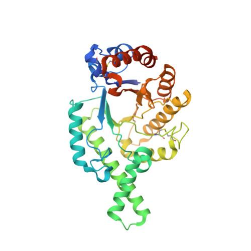
Crystal structure of hyaluronidase, a major allergen of bee venom.
Markovic-Housley, Z., Miglierini, G., Soldatova, L., Rizkallah, P.J., Muller, U., Schirmer, T.(2000) Structure 8: 1025-1035
- PubMed: 11080624
- DOI: https://doi.org/10.1016/s0969-2126(00)00511-6
- Primary Citation of Related Structures:
1FCQ, 1FCU, 1FCV - PubMed Abstract:
Hyaluronic acid (HA) is the most abundant glycosaminoglycan of vertebrate extracellular spaces and is specifically degraded by a beta-1,4 glycosidase. Bee venom hyaluronidase (Hya) shares 30% sequence identity with human hyaluronidases, which are involved in fertilization and the turnover of HA. On the basis of sequence similarity, mammalian enzymes and Hya are assigned to glycosidase family 56 for which no structure has been reported yet. The crystal structure of recombinant (Baculovirus) Hya was determined at 1.6 A resolution. The overall topology resembles a classical (beta/alpha)(8) TIM barrel except that the barrel is composed of only seven strands. A long substrate binding groove extends across the C-terminal end of the barrel. Cocrystallization with a substrate analog revealed the presence of a HA tetramer bound to subsites -4 to -1 and distortion of the -1 sugar. The structure of the complex strongly suggest an acid-base catalytic mechanism, in which Glu113 acts as the proton donor and the N-acetyl group of the substrate is the nucleophile. The location of the catalytic residues shows striking similarity to bacterial chitinase which also operates via a substrate-assisted mechanism.
- Division of Structural Biology Biozentrum University of Basel CH-4056, Basel, Switzerland. zora.housley@unibas.ch
Organizational Affiliation:


