1HTH | pdb_00001hth

The solution structure of cyclic human parathyroid hormone fragment 1-34, NMR, 10 structures


Experimental Data Snapshot

  • Method: SOLUTION NMR
  • Conformers Calculated: 50 
  • Conformers Submitted: 10 
  • Selection Criteria: ENERGY, AGREEMENT WITH EXPERIMENTAL DATA 

wwPDB Validation 3D Report Full Report

Validation slider image for 1HTH

This is version 1.3 of the entry. See complete history

Literature

The structure of human parathyroid hormone-related protein(1-34) in near-physiological solution.

Weidler, M.Marx, U.C.Seidel, G.Schaefer, W.Hoffmann, E.Esswein, A.Roesch, P.

(1999) FEBS Lett 444: 239-244

  • DOI: https://doi.org/10.1016/s0014-5793(98)01658-5
  • Primary Citation Related Structures: 
    1BZG, 1HTH

  • PubMed Abstract: 

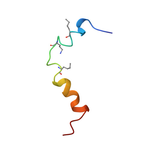
    Parathyroid hormone-related protein plays a major role in the pathogenesis of humoral hypercalcemia of malignancy. Under normal physiological conditions, parathyroid hormone-related protein is produced in a wide variety of tissues and acts in an autocrine or paracrine fashion. Parathyroid hormone-related protein and parathyroid hormone bind to and activate the same G-protein-coupled receptor. Here we present the structure of the biologically active NH2-terminal domain of human parathyroid hormone-related protein(1-34) in near-physiological solution in the absence of crowding reagents as determined by two-dimensional proton magnetic resonance spectroscopy. An improved strategy for structure calculation revealed the presence of two helices, His-5-Leu-8 and Gln-16-Leu-27, connected by a flexible linker. The parathyroid hormone-related protein(1-34) structure and the structure of human parathyroid hormone(1-37) as well as human parathyroid hormone(1-34) are highly similar, except for the well defined turn, His-14-Ser-17, present in parathyroid hormone. Thus, the similarity of the binding affinities of parathyroid hormone and parathyroid hormone-related protein to their common receptor may be based on their structural similarity.


  • Organizational Affiliation
    • Lehrstuhl für Biopolymere, Universität Bayreuth, Germany.

Macromolecule Content 

  • Total Structure Weight: 4.12 kDa 
  • Atom Count: 290 
  • Modeled Residue Count: 34 
  • Deposited Residue Count: 34 
  • Unique protein chains: 1

Macromolecules

Find similar proteins by:|  3D Structure
Entity ID: 1
MoleculeChains  Sequence LengthOrganismDetailsImage
CYCLIC PARATHYROID HORMONE34Homo sapiensMutation(s): 4 
UniProt & NIH Common Fund Data Resources
Find proteins for P01270 (Homo sapiens)
Explore P01270 
Go to UniProtKB:  P01270
PHAROS:  P01270
GTEx:  ENSG00000152266 
Entity Groups
Sequence Clusters30% Identity50% Identity70% Identity90% Identity95% Identity100% Identity
UniProt GroupP01270
Sequence Annotations
Expand
Reference Sequence

Small Molecules

Modified Residues  2 Unique
IDChains TypeFormula2D DiagramParent
NLE
Query on NLE
A
L-PEPTIDE LINKINGC6 H13 N O2LEU
ORN
Query on ORN
A
L-PEPTIDE LINKINGC5 H12 N2 O2ALA

Experimental Data & Validation

Experimental Data

  • Method: SOLUTION NMR
  • Conformers Calculated: 50 
  • Conformers Submitted: 10 
  • Selection Criteria: ENERGY, AGREEMENT WITH EXPERIMENTAL DATA 

Structure Validation

View Full Validation Report


Entry History 

Deposition Data

Revision History  (Full details and data files)

  • Version 1.0: 1997-10-15
    Type: Initial release
  • Version 1.1: 2008-03-24
    Changes: Version format compliance
  • Version 1.2: 2011-07-13
    Changes: Non-polymer description, Version format compliance
  • Version 1.3: 2021-11-03
    Changes: Database references, Derived calculations, Other