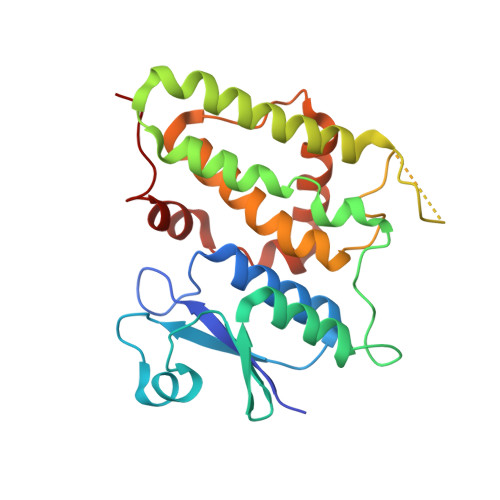
1K0N

Chloride Intracellular Channel 1 (CLIC1) complexed with glutathione


Experimental Data Snapshot

  • Method: X-RAY DIFFRACTION
  • Resolution: 1.80 Å
  • R-Value Free: 0.281 
  • R-Value Work: 0.251 
  • R-Value Observed: 0.252 

wwPDB Validation   3D Report Full Report


This is version 1.5 of the entry. See complete history


Literature

Crystal structure of a soluble form of the intracellular chloride ion channel CLIC1 (NCC27) at 1.4-A resolution.

Harrop, S.J.DeMaere, M.Z.Fairlie, W.D.Reztsova, T.Valenzuela, S.M.Mazzanti, M.Tonini, R.Qiu, M.R.Jankova, L.Warton, K.Bauskin, A.R.Wu, W.M.Pankhurst, S.Campbell, T.J.Breit, S.N.Curmi, P.M.

(2001) J Biol Chem 276: 44993-45000

  • DOI: https://doi.org/10.1074/jbc.M107804200
  • Primary Citation of Related Structures:  
    1K0M, 1K0N, 1K0O

  • PubMed Abstract: 

    CLIC1 (NCC27) is a member of the highly conserved class of chloride ion channels that exists in both soluble and integral membrane forms. Purified CLIC1 can integrate into synthetic lipid bilayers forming a chloride channel with similar properties to those observed in vivo. The structure of the soluble form of CLIC1 has been determined at 1.4-A resolution. The protein is monomeric and structurally homologous to the glutathione S-transferase superfamily, and it has a redox-active site resembling glutaredoxin. The structure of the complex of CLIC1 with glutathione shows that glutathione occupies the redox-active site, which is adjacent to an open, elongated slot lined by basic residues. Integration of CLIC1 into the membrane is likely to require a major structural rearrangement, probably of the N-domain (residues 1-90), with the putative transmembrane helix arising from residues in the vicinity of the redox-active site. The structure indicates that CLIC1 is likely to be controlled by redox-dependent processes.


  • Organizational Affiliation

    Initiative for Biomolecular Structure, School of Physics and the Department of Medicine, University of New South Wales, New South Wales 2052, Australia.


Macromolecules
Find similar proteins by:  (by identity cutoff)  |  3D Structure
Entity ID: 1
MoleculeChains Sequence LengthOrganismDetailsImage
CHLORIDE INTRACELLULAR CHANNEL PROTEIN 1
A, B
241Homo sapiensMutation(s): 1 
EC: 1.8 (UniProt), 1.8.5.1 (UniProt)
UniProt & NIH Common Fund Data Resources
Find proteins for O00299 (Homo sapiens)
Explore O00299 
Go to UniProtKB:  O00299
PHAROS:  O00299
GTEx:  ENSG00000213719 
Entity Groups  
Sequence Clusters30% Identity50% Identity70% Identity90% Identity95% Identity100% Identity
UniProt GroupO00299
Sequence Annotations
Expand
  • Reference Sequence
Small Molecules
Ligands 1 Unique
IDChains Name / Formula / InChI Key2D Diagram3D Interactions
GSH
Query on GSH

Download Ideal Coordinates CCD File 
C [auth A]GLUTATHIONE
C10 H17 N3 O6 S
RWSXRVCMGQZWBV-WDSKDSINSA-N
Experimental Data & Validation

Experimental Data

  • Method: X-RAY DIFFRACTION
  • Resolution: 1.80 Å
  • R-Value Free: 0.281 
  • R-Value Work: 0.251 
  • R-Value Observed: 0.252 
  • Space Group: P 1 21 1
Unit Cell:
Length ( Å )Angle ( ˚ )
a = 46.509α = 90
b = 60.355β = 92.5
c = 89.627γ = 90
Software Package:
Software NamePurpose
AMoREphasing
REFMACrefinement
DENZOdata reduction
SCALEPACKdata scaling

Structure Validation

View Full Validation Report


Entry History 

Revision History  (Full details and data files)

  • Version 1.0: 2001-12-12
    Type: Initial release
  • Version 1.1: 2008-04-27
    Changes: Version format compliance
  • Version 1.2: 2011-07-13
    Changes: Version format compliance
  • Version 1.3: 2011-12-21
    Changes: Non-polymer description
  • Version 1.4: 2021-10-27
    Changes: Database references, Derived calculations
  • Version 1.5: 2024-02-07
    Changes: Data collection