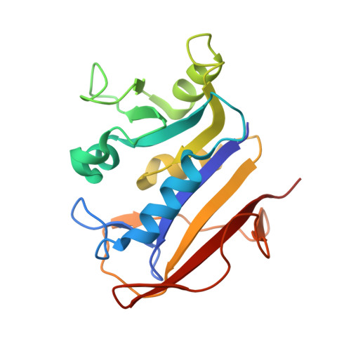
1KMS

HUMAN DIHYDROFOLATE REDUCTASE COMPLEXED WITH NADPH AND 6-([5-QUINOLYLAMINO]METHYL)-2,4-DIAMINO-5-METHYLPYRIDO[2,3-D]PYRIMIDINE (SRI-9439), A LIPOPHILIC ANTIFOLATE


Experimental Data Snapshot

  • Method: X-RAY DIFFRACTION
  • Resolution: 1.09 Å
  • R-Value Free: 0.173 
  • R-Value Work: 0.131 

Starting Model: other
View more details

wwPDB Validation   3D Report Full Report


Ligand Structure Quality Assessment 


This is version 1.4 of the entry. See complete history


Literature

Atomic structures of human dihydrofolate reductase complexed with NADPH and two lipophilic antifolates at 1.09 a and 1.05 a resolution.

Klon, A.E.Heroux, A.Ross, L.J.Pathak, V.Johnson, C.A.Piper, J.R.Borhani, D.W.

(2002) J Mol Biol 320: 677-693

  • DOI: https://doi.org/10.1016/s0022-2836(02)00469-2
  • Primary Citation of Related Structures:  
    1KMS, 1KMV

  • PubMed Abstract: 

    The crystal structures of two human dihydrofolate reductase (hDHFR) ternary complexes, each with bound NADPH cofactor and a lipophilic antifolate inhibitor, have been determined at atomic resolution. The potent inhibitors 6-([5-quinolylamino]methyl)-2,4-diamino-5-methylpyrido[2,3-d]pyrimidine (SRI-9439) and (Z)-6-(2-[2,5-dimethoxyphenyl]ethen-1-yl)-2,4-diamino-5-methylpyrido[2,3-d]pyrimidine (SRI-9662) were developed at Southern Research Institute against Toxoplasma gondii DHFR-thymidylate synthase. The 5-deazapteridine ring of each inhibitor adopts an unusual puckered conformation that enables the formation of identical contacts in the active site. Conversely, the quinoline and dimethoxybenzene moieties exhibit distinct binding characteristics that account for the differences in inhibitory activity. In both structures, a salt-bridge is formed between Arg70 in the active site and Glu44 from a symmetry-related molecule in the crystal lattice that mimics the binding of methotrexate to DHFR.


  • Organizational Affiliation

    Department of Biochemistry and Molecular Genetics, University of Alabama at Birmingham, 35294, USA.


Macromolecules
Find similar proteins by:  (by identity cutoff)  |  3D Structure
Entity ID: 1
MoleculeChains Sequence LengthOrganismDetailsImage
DIHYDROFOLATE REDUCTASE186Homo sapiensMutation(s): 0 
EC: 1.5.1.3
UniProt & NIH Common Fund Data Resources
Find proteins for P00374 (Homo sapiens)
Explore P00374 
Go to UniProtKB:  P00374
PHAROS:  P00374
GTEx:  ENSG00000228716 
Entity Groups  
Sequence Clusters30% Identity50% Identity70% Identity90% Identity95% Identity100% Identity
UniProt GroupP00374
Sequence Annotations
Expand
  • Reference Sequence
Small Molecules
Binding Affinity Annotations 
IDSourceBinding Affinity
LIH BindingDB:  1KMS IC50: min: 0.98, max: 4.3 (nM) from 2 assay(s)
Experimental Data & Validation

Experimental Data

  • Method: X-RAY DIFFRACTION
  • Resolution: 1.09 Å
  • R-Value Free: 0.173 
  • R-Value Work: 0.131 
  • Space Group: P 21 21 21
Unit Cell:
Length ( Å )Angle ( ˚ )
a = 41.467α = 90
b = 55.275β = 90
c = 82.799γ = 90
Software Package:
Software NamePurpose
AMoREphasing
SHELXL-97refinement
MOSFLMdata reduction
CCP4data scaling

Structure Validation

View Full Validation Report


Ligand Structure Quality Assessment 


Entry History 

Deposition Data

Revision History  (Full details and data files)

  • Version 1.0: 2002-07-10
    Type: Initial release
  • Version 1.1: 2008-04-27
    Changes: Version format compliance
  • Version 1.2: 2011-07-13
    Changes: Version format compliance
  • Version 1.3: 2024-02-14
    Changes: Data collection, Database references, Derived calculations
  • Version 1.4: 2024-04-03
    Changes: Refinement description