1MK2 | pdb_00001mk2

SMAD3 SBD complex


Experimental Data Snapshot

  • Method: X-RAY DIFFRACTION
  • Resolution: 2.74 Å
  • R-Value Free: 
    0.271 (Depositor), 0.266 (DCC) 
  • R-Value Work: 
    0.227 (Depositor), 0.227 (DCC) 
  • R-Value Observed: 
    0.227 (Depositor) 

wwPDB Validation 3D Report Full Report

Validation slider image for 1MK2

This is version 1.3 of the entry. See complete history

Literature

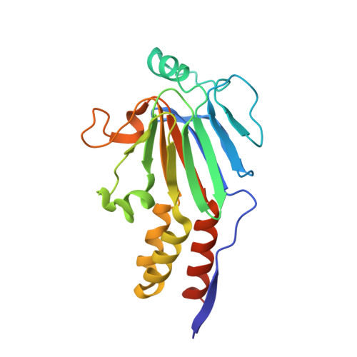
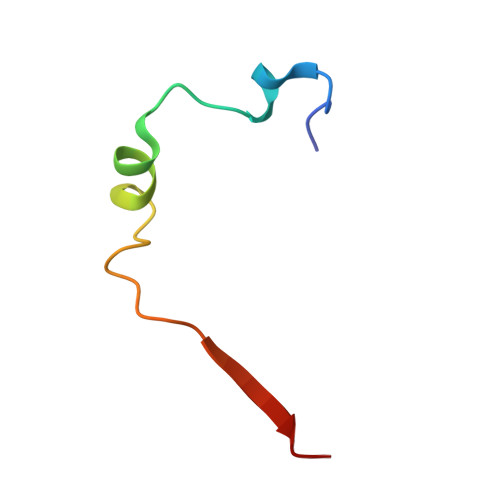
Smad3 allostery links TGF-beta receptor kinase activation to transcriptional control

Qin, B.Y.Lam, S.S.Correia, J.J.Lin, K.

(2002) Genes Dev 16: 1950-1963

  • DOI: https://doi.org/10.1101/gad.1002002
  • Primary Citation Related Structures: 
    1MJS, 1MK2

  • PubMed Abstract: 

    Smad3 transduces the signals of TGF-betas, coupling transmembrane receptor kinase activation to transcriptional control. The membrane-associated molecule SARA (Smad Anchor for Receptor Activation) recruits Smad3 for phosphorylation by the receptor kinase. Upon phosphorylation, Smad3 dissociates from SARA and enters the nucleus, in which its transcriptional activity can be repressed by Ski. Here, we show that SARA and Ski recognize specifically the monomeric and trimeric forms of Smad3, respectively. Thus, trimerization of Smad3, induced by phosphorylation, simultaneously activates the TGF-beta signal by driving Smad3 dissociation from SARA and sets up the negative feedback mechanism by Ski. Structural models of the Smad3/SARA/receptor kinase complex and Smad3/Ski complex provide insights into the molecular basis of regulation.


  • Organizational Affiliation
    • Department of Biochemistry and Molecular Pharmacology, University of Massachusetts Medical School, Worcester, Massachusetts 01655, USA.

Macromolecule Content 

  • Total Structure Weight: 27.29 kDa 
  • Atom Count: 1,848 
  • Modeled Residue Count: 235 
  • Deposited Residue Count: 244 
  • Unique protein chains: 2

Macromolecules

Find similar proteins by:|  3D Structure
Entity ID: 1
MoleculeChains  Sequence LengthOrganismDetailsImage
SMAD 3206Homo sapiensMutation(s): 0 
UniProt & NIH Common Fund Data Resources
Find proteins for P84022 (Homo sapiens)
Explore P84022 
Go to UniProtKB:  P84022
PHAROS:  P84022
GTEx:  ENSG00000166949 
Entity Groups
Sequence Clusters30% Identity50% Identity70% Identity90% Identity95% Identity100% Identity
UniProt GroupP84022
Sequence Annotations
Expand
Reference Sequence
Find similar proteins by:|  3D Structure
Entity ID: 2
MoleculeChains  Sequence LengthOrganismDetailsImage
Madh-interacting protein38Homo sapiensMutation(s): 0 
UniProt & NIH Common Fund Data Resources
Find proteins for O95405 (Homo sapiens)
Explore O95405 
Go to UniProtKB:  O95405
PHAROS:  O95405
GTEx:  ENSG00000157077 
Entity Groups
Sequence Clusters30% Identity50% Identity70% Identity90% Identity95% Identity100% Identity
UniProt GroupO95405
Sequence Annotations
Expand
Reference Sequence

Experimental Data & Validation

Experimental Data

  • Method: X-RAY DIFFRACTION
  • Resolution: 2.74 Å
  • R-Value Free:  0.271 (Depositor), 0.266 (DCC) 
  • R-Value Work:  0.227 (Depositor), 0.227 (DCC) 
  • R-Value Observed: 0.227 (Depositor) 
Space Group: P 21 21 2
Unit Cell:
Length ( Å )Angle ( ˚ )
a = 49.995α = 90
b = 71.716β = 90
c = 86.886γ = 90
Software Package:
Software NamePurpose
DENZOdata reduction
SCALEPACKdata scaling
CNSrefinement
CNSphasing

Structure Validation

View Full Validation Report


Entry History 

Deposition Data

Revision History  (Full details and data files)

  • Version 1.0: 2002-10-16
    Type: Initial release
  • Version 1.1: 2008-04-28
    Changes: Version format compliance
  • Version 1.2: 2011-07-13
    Changes: Derived calculations, Version format compliance
  • Version 1.3: 2024-02-14
    Changes: Data collection, Database references, Derived calculations