1MRO | pdb_00001mro

METHYL-COENZYME M REDUCTASE


Experimental Data Snapshot

  • Method: X-RAY DIFFRACTION
  • Resolution: 1.16 Å
  • R-Value Free: 
    0.207 (Depositor) 
  • R-Value Work: 
    0.197 (Depositor) 
  • R-Value Observed: 
    0.197 (Depositor) 

wwPDB Validation 3D Report Full Report

Validation slider image for 1MRO

This is version 1.6 of the entry. See complete history

Literature

Crystal structure of methyl-coenzyme M reductase: the key enzyme of biological methane formation.

Ermler, U.Grabarse, W.Shima, S.Goubeaud, M.Thauer, R.K.

(1997) Science 278: 1457-1462

  • DOI: https://doi.org/10.1126/science.278.5342.1457
  • Primary Citation Related Structures: 
    1MRO

  • PubMed Abstract: 

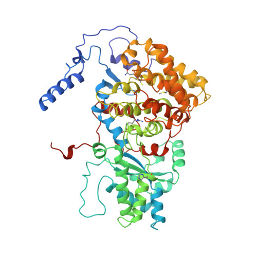
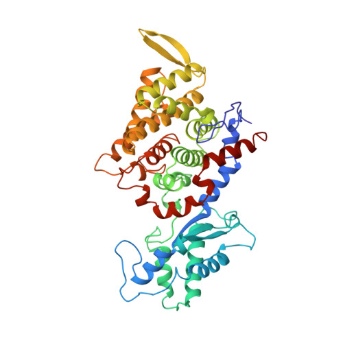
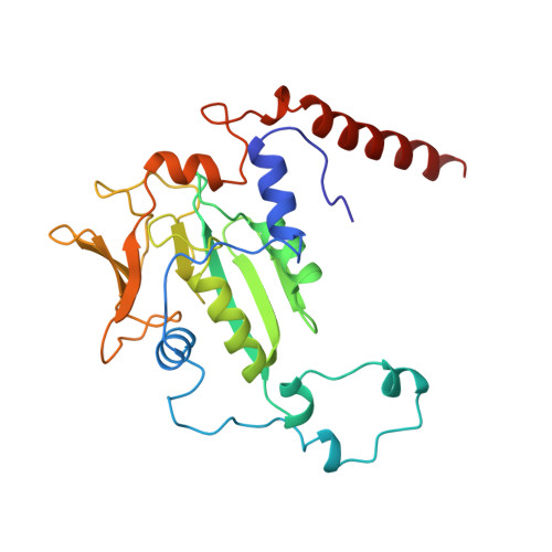
    Methyl-coenzyme M reductase (MCR), the enzyme responsible for the microbial formation of methane, is a 300-kilodalton protein organized as a hexamer in an alpha2beta2gamma2 arrangement. The crystal structure of the enzyme from Methanobacterium thermoautotrophicum, determined at 1.45 angstrom resolution for the inactive enzyme state MCRox1-silent, reveals that two molecules of the nickel porphinoid coenzyme F430 are embedded between the subunits alpha, alpha', beta, and gamma and alpha', alpha, beta', and gamma', forming two identical active sites. Each site is accessible for the substrate methyl-coenzyme M through a narrow channel locked after binding of the second substrate coenzyme B. Together with a second structurally characterized enzyme state (MCRsilent) containing the heterodisulfide of coenzymes M and B, a reaction mechanism is proposed that uses a radical intermediate and a nickel organic compound.


  • Organizational Affiliation
    • Max-Planck-Institut für Biophysik, Heinrich-Hoffmann-Strabetae 7, 60528 Frankfurt, Germany.

Macromolecule Content 

  • Total Structure Weight: 275.39 kDa 
  • Atom Count: 21,217 
  • Modeled Residue Count: 2,474 
  • Deposited Residue Count: 2,474 
  • Unique protein chains: 3

Macromolecules

Find similar proteins by:|  3D Structure
Entity ID: 1
MoleculeChains  Sequence LengthOrganismDetailsImage
METHYL-COENZYME M REDUCTASE
A, D
548Methanothermobacter marburgensis str. MarburgMutation(s): 0 
EC: 1.8 (PDB Primary Data), 2.8.4.1 (UniProt)
UniProt
Find proteins for P11558 (Methanothermobacter marburgensis (strain ATCC BAA-927 / DSM 2133 / JCM 14651 / NBRC 100331 / OCM 82 / Marburg))
Explore P11558 
Go to UniProtKB:  P11558
Entity Groups
Sequence Clusters30% Identity50% Identity70% Identity90% Identity95% Identity100% Identity
UniProt GroupP11558
Sequence Annotations
Expand
Reference Sequence
Find similar proteins by:|  3D Structure
Entity ID: 2
MoleculeChains  Sequence LengthOrganismDetailsImage
METHYL-COENZYME M REDUCTASE
B, E
442Methanothermobacter marburgensis str. MarburgMutation(s): 0 
EC: 1.8 (PDB Primary Data), 2.8.4.1 (UniProt)
UniProt
Find proteins for P11560 (Methanothermobacter marburgensis (strain ATCC BAA-927 / DSM 2133 / JCM 14651 / NBRC 100331 / OCM 82 / Marburg))
Explore P11560 
Go to UniProtKB:  P11560
Entity Groups
Sequence Clusters30% Identity50% Identity70% Identity90% Identity95% Identity100% Identity
UniProt GroupP11560
Sequence Annotations
Expand
Reference Sequence
Find similar proteins by:|  3D Structure
Entity ID: 3
MoleculeChains  Sequence LengthOrganismDetailsImage
METHYL-COENZYME M REDUCTASE
C, F
247Methanothermobacter marburgensis str. MarburgMutation(s): 0 
EC: 1.8 (PDB Primary Data), 2.8.4.1 (UniProt)
UniProt
Find proteins for P11562 (Methanothermobacter marburgensis (strain ATCC BAA-927 / DSM 2133 / JCM 14651 / NBRC 100331 / OCM 82 / Marburg))
Explore P11562 
Go to UniProtKB:  P11562
Entity Groups
Sequence Clusters30% Identity50% Identity70% Identity90% Identity95% Identity100% Identity
UniProt GroupP11562
Sequence Annotations
Expand
Reference Sequence

Small Molecules

Ligands 6 Unique
IDChains Name / Formula / InChI Key2D Diagram3D Interactions
F43

Query on F43


Download:Ideal Coordinates CCD File
H [auth A],
O [auth D]
FACTOR 430
C42 H51 N6 Ni O13
XLFIRMYGVLUNOY-SXMZNAGASA-M
TP7

Query on TP7


Download:Ideal Coordinates CCD File
I [auth A],
J [auth A]
Coenzyme B
C11 H22 N O7 P S
JBJSVEVEEGOEBZ-SCZZXKLOSA-N
COM

Query on COM


Download:Ideal Coordinates CCD File
K [auth A],
P [auth D]
1-THIOETHANESULFONIC ACID
C2 H6 O3 S2
ZNEWHQLOPFWXOF-UHFFFAOYSA-N
GOL

Query on GOL


Download:Ideal Coordinates CCD File
L [auth A],
M [auth A],
Q [auth D],
R [auth D]
GLYCEROL
C3 H8 O3
PEDCQBHIVMGVHV-UHFFFAOYSA-N
ZN

Query on ZN


Download:Ideal Coordinates CCD File
G [auth A]ZINC ION
Zn
PTFCDOFLOPIGGS-UHFFFAOYSA-N
NA

Query on NA


Download:Ideal Coordinates CCD File
N [auth C],
S [auth F]
SODIUM ION
Na
FKNQFGJONOIPTF-UHFFFAOYSA-N
Modified Residues  5 Unique
IDChains TypeFormula2D DiagramParent
AGM
Query on AGM
A, D
L-PEPTIDE LINKINGC7 H17 N4 O2ARG
GL3
Query on GL3
A, D
L-PEPTIDE LINKINGC2 H5 N O SGLY
MGN
Query on MGN
A, D
L-PEPTIDE LINKINGC6 H12 N2 O3GLN
MHS
Query on MHS
A, D
L-PEPTIDE LINKINGC7 H11 N3 O2HIS
SMC
Query on SMC
A, D
L-PEPTIDE LINKINGC4 H9 N O2 SCYS

Experimental Data & Validation

Experimental Data

  • Method: X-RAY DIFFRACTION
  • Resolution: 1.16 Å
  • R-Value Free:  0.207 (Depositor) 
  • R-Value Work:  0.197 (Depositor) 
  • R-Value Observed: 0.197 (Depositor) 
Space Group: P 1 21 1
Unit Cell:
Length ( Å )Angle ( ˚ )
a = 81.72α = 90
b = 116.883β = 92.02
c = 122.582γ = 90
Software Package:
Software NamePurpose
CNSrefinement
DENZOdata reduction
SCALEPACKdata scaling
CNSphasing

Structure Validation

View Full Validation Report


Entry History 

Deposition Data

Revision History  (Full details and data files)

  • Version 1.0: 1998-11-11
    Type: Initial release
  • Version 1.1: 2008-03-24
    Changes: Version format compliance
  • Version 1.2: 2011-07-13
    Changes: Non-polymer description, Version format compliance
  • Version 1.3: 2015-03-04
    Changes: Derived calculations
  • Version 1.4: 2015-05-20
    Changes: Non-polymer description
  • Version 1.5: 2017-03-29
    Changes: Non-polymer description
  • Version 1.6: 2025-03-26
    Changes: Data collection, Database references, Derived calculations, Structure summary