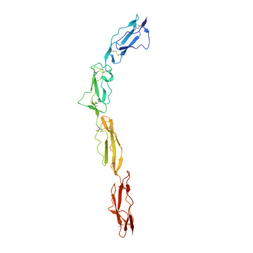
1OJY

Decay accelerating factor (cd55): the structure of an intact human complement regulator.


Experimental Data Snapshot

  • Method: X-RAY DIFFRACTION
  • Resolution: 2.60 Å

Starting Model: experimental
View more details

wwPDB Validation   3D Report Full Report


This is version 1.3 of the entry. See complete history


Literature

Complement Regulation at the Molecular Level: The Structure of Decay-Accelerating Factor

Lukacik, P.Roversi, P.White, J.Esser, D.Smith, G.P.Billington, J.Williams, P.A.Rudd, P.M.Wormald, M.R.Harvey, D.J.Crispin, M.D.M.Radcliffe, C.M.Dwek, R.A.Evans, D.J.Morgan, B.P.Smith, R.A.G.Lea, S.M.

(2004) Proc Natl Acad Sci U S A 101: 1279

  • DOI: https://doi.org/10.1073/pnas.0307200101
  • Primary Citation of Related Structures:  
    1OJV, 1OJW, 1OJY, 1OK1, 1OK2, 1OK3, 1OK9

  • PubMed Abstract: 

    The human complement regulator CD55 is a key molecule protecting self-cells from complement-mediated lysis. X-ray diffraction and analytical ultracentrifugation data reveal a rod-like arrangement of four short consensus repeat (SCR) domains in both the crystal and solution. The stalk linking the four SCR domains to the glycosylphosphatidylinositol anchor is extended by the addition of 11 highly charged O-glycans and positions the domains an estimated 177 A above the membrane. Mutation mapping and hydrophobic potential analysis suggest that the interaction with the convertase, and thus complement regulation, depends on the burial of a hydrophobic patch centered on the linker between SCR domains 2 and 3.


  • Organizational Affiliation

    Laboratory of Molecular Biophysics and Glycobiology Institute, Department of Biochemistry, University of Oxford, South Parks Road, Oxford OX1 3QU, England.


Macromolecules
Find similar proteins by:  (by identity cutoff)  |  3D Structure
Entity ID: 1
MoleculeChains Sequence LengthOrganismDetailsImage
COMPLEMENT DECAY-ACCELERATING FACTOR
A, B, C, D
254Homo sapiensMutation(s): 0 
UniProt & NIH Common Fund Data Resources
Find proteins for P08174 (Homo sapiens)
Explore P08174 
Go to UniProtKB:  P08174
PHAROS:  P08174
GTEx:  ENSG00000196352 
Entity Groups  
Sequence Clusters30% Identity50% Identity70% Identity90% Identity95% Identity100% Identity
UniProt GroupP08174
Sequence Annotations
Expand
  • Reference Sequence
Small Molecules
Ligands 3 Unique
IDChains Name / Formula / InChI Key2D Diagram3D Interactions
SO4
Query on SO4

Download Ideal Coordinates CCD File 
H [auth C],
K [auth C],
M [auth D]
SULFATE ION
O4 S
QAOWNCQODCNURD-UHFFFAOYSA-L
GOL
Query on GOL

Download Ideal Coordinates CCD File 
G [auth B]
I [auth C]
L [auth C]
O [auth D]
P [auth D]
G [auth B],
I [auth C],
L [auth C],
O [auth D],
P [auth D],
Q [auth D]
GLYCEROL
C3 H8 O3
PEDCQBHIVMGVHV-UHFFFAOYSA-N
ACT
Query on ACT

Download Ideal Coordinates CCD File 
E [auth A],
F [auth B],
J [auth C],
N [auth D]
ACETATE ION
C2 H3 O2
QTBSBXVTEAMEQO-UHFFFAOYSA-M
Experimental Data & Validation

Experimental Data

  • Method: X-RAY DIFFRACTION
  • Resolution: 2.60 Å
  • Space Group: P 1
Unit Cell:
Length ( Å )Angle ( ˚ )
a = 51.829α = 72.7
b = 78.694β = 79.38
c = 80.903γ = 81.7
Software Package:
Software NamePurpose
TNTrefinement
DENZOdata reduction
SCALEPACKdata scaling
MOLREPphasing

Structure Validation

View Full Validation Report


Entry History 

Revision History  (Full details and data files)

  • Version 1.0: 2004-01-07
    Type: Initial release
  • Version 1.1: 2016-09-21
    Changes: Database references, Derived calculations, Non-polymer description, Other, Refinement description, Structure summary, Version format compliance
  • Version 1.2: 2023-12-13
    Changes: Data collection, Database references, Derived calculations, Other, Refinement description
  • Version 1.3: 2024-10-09
    Changes: Structure summary