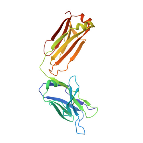
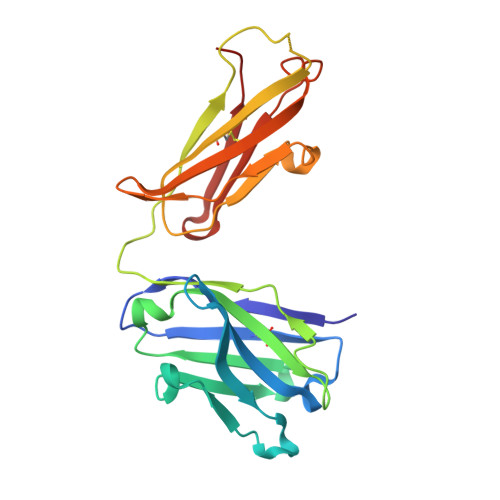
1QYG

ANTI-COCAINE ANTIBODY M82G2 COMPLEXED WITH BENZOYLECGONINE


Experimental Data Snapshot

  • Method: X-RAY DIFFRACTION
  • Resolution: 1.81 Å
  • R-Value Free: 0.216 
  • R-Value Work: 0.178 
  • R-Value Observed: 0.178 

Starting Models: experimental
View more details

wwPDB Validation   3D Report Full Report


Ligand Structure Quality Assessment 


This is version 1.4 of the entry. See complete history


Literature

Diversity in hapten recognition: structural study of an anti-cocaine antibody M82G2.

Pozharski, E.Moulin, A.Hewagama, A.Shanafelt, A.B.Petsko, G.A.Ringe, D.

(2005) J Mol Biol 349: 570-582

  • DOI: https://doi.org/10.1016/j.jmb.2005.03.080
  • Primary Citation of Related Structures:  
    1Q72, 1QYG, 1RFD

  • PubMed Abstract: 

    Antibodies against cocaine and other drugs of abuse are the basis for diagnostic tests for the presence of those drugs in human serum. The 1.7A resolution crystal structure of the anti-cocaine monoclonal antibody M82G2 in complex with cocaine is presented. This structure determination was undertaken to establish the stereochemical features in the antibody binding site that confer specificity for cocaine, and as part of an ongoing project to understand the rules that govern molecular recognition. The cocaine-binding site can be characterized topologically as a narrow groove on the protein surface. The antibody utilizes water-mediated hydrogen bonding, and cation-pi and stacking (pi-pi) interactions to provide specificity. Comparison with the previously published structure of the anti-cocaine antibody GNC92H2 shows that binding of a small ligand can be achieved in diverse ways, both in terms of a binding site structure/topology and protein-ligand interactions.


  • Organizational Affiliation

    Rosenstiel Basic Medical Sciences Research Center, Brandeis University, 415 South Street, Waltham, MA 02454, USA.


Macromolecules
Find similar proteins by:  (by identity cutoff)  |  3D Structure
Entity ID: 1
MoleculeChains Sequence LengthOrganismDetailsImage
FAB M82G2, LIGHT CHAINA [auth L]218Mus musculusMutation(s): 0 
Entity Groups  
Sequence Clusters30% Identity50% Identity70% Identity90% Identity95% Identity100% Identity
Sequence Annotations
Expand
  • Reference Sequence
Find similar proteins by:  (by identity cutoff)  |  3D Structure
Entity ID: 2
MoleculeChains Sequence LengthOrganismDetailsImage
FAB M82G2, HEAVY CHAINB [auth H]223Mus musculusMutation(s): 0 
Entity Groups  
Sequence Clusters30% Identity50% Identity70% Identity90% Identity95% Identity100% Identity
Sequence Annotations
Expand
  • Reference Sequence
Small Molecules
Ligands 1 Unique
IDChains Name / Formula / InChI Key2D Diagram3D Interactions
BCG
Query on BCG

Download Ideal Coordinates CCD File 
C [auth H]3-(BENZOYLOXY)-8-METHYL-8-AZABICYCLO[3.2.1]OCTANE-2-CARBOXYLIC ACID
C16 H19 N O4
GVGYEFKIHJTNQZ-RFQIPJPRSA-N
Binding Affinity Annotations 
IDSourceBinding Affinity
BCG PDBBind:  1QYG Kd: 140 (nM) from 1 assay(s)
Experimental Data & Validation

Experimental Data

  • Method: X-RAY DIFFRACTION
  • Resolution: 1.81 Å
  • R-Value Free: 0.216 
  • R-Value Work: 0.178 
  • R-Value Observed: 0.178 
  • Space Group: P 31 2 1
Unit Cell:
Length ( Å )Angle ( ˚ )
a = 75.17α = 90
b = 75.17β = 90
c = 151.276γ = 120
Software Package:
Software NamePurpose
DENZOdata reduction
SCALEPACKdata scaling
AMoREphasing
CNSrefinement

Structure Validation

View Full Validation Report


Ligand Structure Quality Assessment 


Entry History 

Deposition Data

Revision History  (Full details and data files)

  • Version 1.0: 2003-09-23
    Type: Initial release
  • Version 1.1: 2008-04-29
    Changes: Version format compliance
  • Version 1.2: 2011-07-13
    Changes: Version format compliance
  • Version 1.3: 2023-08-23
    Changes: Data collection, Database references, Derived calculations, Refinement description
  • Version 1.4: 2024-11-06
    Changes: Structure summary