1SHY | pdb_00001shy

The Crystal Structure of HGF beta-chain in Complex with the Sema Domain of the Met Receptor.


Experimental Data Snapshot

  • Method: X-RAY DIFFRACTION
  • Resolution: 3.22 Å
  • R-Value Free: 
    0.270 (Depositor), 0.248 (DCC) 
  • R-Value Work: 
    0.209 (Depositor), 0.197 (DCC) 
  • R-Value Observed: 
    0.212 (Depositor) 

Starting Model: other
View more details

wwPDB Validation 3D Report Full Report

Validation slider image for 1SHY

This is version 1.5 of the entry. See complete history

Literature

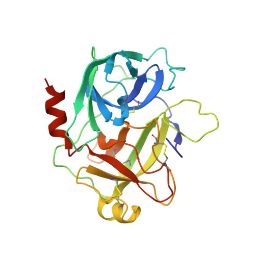
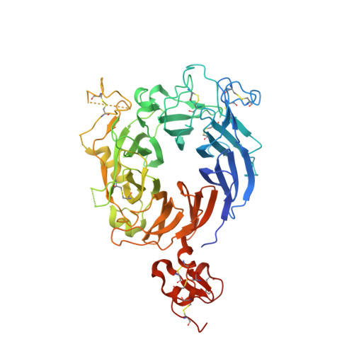
Crystal structure of the HGF beta-chain in complex with the Sema domain of the Met receptor.

Stamos, J.Lazarus, R.A.Yao, X.Kirchhofer, D.Wiesmann, C.

(2004) EMBO J 23: 2325-2335

  • DOI: https://doi.org/10.1038/sj.emboj.7600243
  • Primary Citation Related Structures: 
    1SHY

  • PubMed Abstract: 

    The Met tyrosine kinase receptor and its ligand, hepatocyte growth factor (HGF), play important roles in normal development and in tumor growth and metastasis. HGF-dependent signaling requires proteolysis from an inactive single-chain precursor into an active alpha/beta-heterodimer. We show that the serine protease-like HGF beta-chain alone binds Met, and report its crystal structure in complex with the Sema and PSI domain of the Met receptor. The Met Sema domain folds into a seven-bladed beta-propeller, where the bottom face of blades 2 and 3 binds to the HGF beta-chain 'active site region'. Mutation of HGF residues in the area that constitutes the active site region in related serine proteases significantly impairs HGF beta binding to Met. Key binding loops in this interface undergo conformational rearrangements upon maturation and explain the necessity of proteolytic cleavage for proper HGF signaling. A crystallographic dimer interface between two HGF beta-chains brings two HGF beta:Met complexes together, suggesting a possible mechanism of Met receptor dimerization and activation by HGF.


  • Organizational Affiliation
    • Department of Protein Engineering, Genentech Inc., South San Francisco, CA 94080, USA.

Macromolecule Content 

  • Total Structure Weight: 88.55 kDa 
  • Atom Count: 5,735 
  • Modeled Residue Count: 727 
  • Deposited Residue Count: 785 
  • Unique protein chains: 2

Macromolecules

Find similar proteins by:|  3D Structure
Entity ID: 1
MoleculeChains  Sequence LengthOrganismDetailsImage
Hepatocyte growth factor234Homo sapiensMutation(s): 1 
Gene Names: HGFHPTA
UniProt & NIH Common Fund Data Resources
Find proteins for P14210 (Homo sapiens)
Explore P14210 
Go to UniProtKB:  P14210
PHAROS:  P14210
GTEx:  ENSG00000019991 
Entity Groups
Sequence Clusters30% Identity50% Identity70% Identity90% Identity95% Identity100% Identity
UniProt GroupP14210
Sequence Annotations
Expand
Reference Sequence
Find similar proteins by:|  3D Structure
Entity ID: 2
MoleculeChains  Sequence LengthOrganismDetailsImage
Hepatocyte growth factor receptor551Homo sapiensMutation(s): 0 
Gene Names: MET
EC: 2.7.10.1
UniProt & NIH Common Fund Data Resources
Find proteins for P08581 (Homo sapiens)
Explore P08581 
Go to UniProtKB:  P08581
PHAROS:  P08581
GTEx:  ENSG00000105976 
Entity Groups
Sequence Clusters30% Identity50% Identity70% Identity90% Identity95% Identity100% Identity
UniProt GroupP08581
Sequence Annotations
Expand
Reference Sequence

Experimental Data & Validation

Experimental Data

  • Method: X-RAY DIFFRACTION
  • Resolution: 3.22 Å
  • R-Value Free:  0.270 (Depositor), 0.248 (DCC) 
  • R-Value Work:  0.209 (Depositor), 0.197 (DCC) 
  • R-Value Observed: 0.212 (Depositor) 
Space Group: P 21 21 2
Unit Cell:
Length ( Å )Angle ( ˚ )
a = 137.052α = 90
b = 186.352β = 90
c = 66.659γ = 90
Software Package:
Software NamePurpose
REFMACrefinement
HKL-2000data reduction
SCALEPACKdata scaling
AMoREphasing

Structure Validation

View Full Validation Report


Entry History 

Deposition Data

Revision History  (Full details and data files)

  • Version 1.0: 2004-06-15
    Type: Initial release
  • Version 1.1: 2008-04-29
    Changes: Version format compliance
  • Version 1.2: 2011-07-13
    Changes: Advisory, Version format compliance
  • Version 1.3: 2021-10-27
    Changes: Database references
  • Version 1.4: 2024-04-03
    Changes: Data collection, Refinement description
  • Version 1.5: 2024-11-13
    Changes: Structure summary