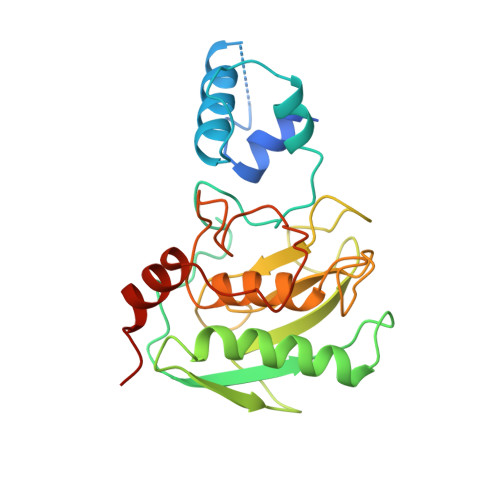
Stromelysin-1: three-dimensional structure of the inhibited catalytic domain and of the C-truncated proenzyme.
Becker, J.W., Marcy, A.I., Rokosz, L.L., Axel, M.G., Burbaum, J.J., Fitzgerald, P.M., Cameron, P.M., Esser, C.K., Hagmann, W.K., Hermes, J.D., Springer, J.P.(1995) Protein Sci 4: 1966-1976
- PubMed: 8535233
- DOI: https://doi.org/10.1002/pro.5560041002
- Primary Citation of Related Structures:
1SLM, 1SLN - PubMed Abstract:
The proteolytic enzyme stromelysin-1 is a member of the family of matrix metalloproteinases and is believed to play a role in pathological conditions such as arthritis and tumor invasion. Stromelysin-1 is synthesized as a pro-enzyme that is activated by removal of an N-terminal prodomain. The active enzyme contains a catalytic domain and a C-terminal hemopexin domain believed to participate in macromolecular substrate recognition. We have determined the three-dimensional structures of both a C-truncated form of the proenzyme and an inhibited complex of the catalytic domain by X-ray diffraction analysis. The catalytic core is very similar in the two forms and is similar to the homologous domain in fibroblast and neutrophil collagenases, as well as to the stromelysin structure determined by NMR. The prodomain is a separate folding unit containing three alpha-helices and an extended peptide that lies in the active site of the enzyme. Surprisingly, the amino-to-carboxyl direction of this peptide chain is opposite to that adopted by the inhibitor and by previously reported inhibitors of collagenase. Comparison of the active site of stromelysin with that of thermolysin reveals that most of the residues proposed to play significant roles in the enzymatic mechanism of thermolysin have equivalents in stromelysin, but that three residues implicated in the catalytic mechanism of thermolysin are not represented in stromelysin.
- Department of Molecular Design and Diversity, Merck Research Laboratories, Rahway, New Jersey 07065, USA.
Organizational Affiliation:


