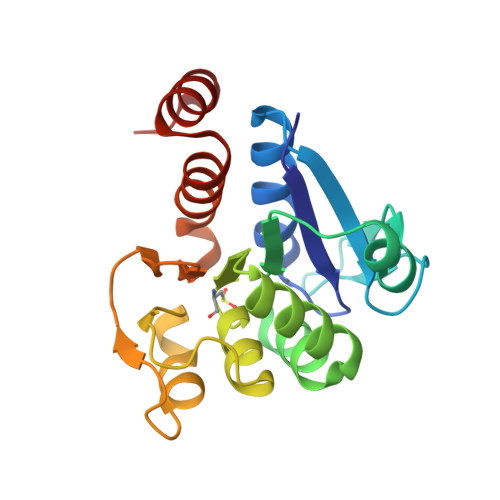
1SOA

Human DJ-1 with sulfinic acid


Experimental Data Snapshot

  • Method: X-RAY DIFFRACTION
  • Resolution: 1.20 Å
  • R-Value Free: 0.141 
  • R-Value Work: 0.113 
  • R-Value Observed: 0.113 

Starting Model: experimental
View more details

wwPDB Validation   3D Report Full Report


This is version 1.6 of the entry. See complete history


Literature

The Parkinson's disease protein DJ-1 is neuroprotective due to cysteine-sulfinic acid-driven mitochondrial localization.

Wilson, M.A.Miller, D.W.Ahmad, R.McLendon, C.Bandyopadhyay, S.Baptista, M.J.Ringe, D.Petsko, G.A.Cookson, M.R.

(2004) Proc Natl Acad Sci U S A 101: 9103-9108

  • DOI: https://doi.org/10.1073/pnas.0402959101
  • Primary Citation of Related Structures:  
    1SOA

  • PubMed Abstract: 

    Loss-of-function DJ-1 mutations can cause early-onset Parkinson's disease. The function of DJ-1 is unknown, but an acidic isoform accumulates after oxidative stress, leading to the suggestion that DJ-1 is protective under these conditions. We addressed whether this represents a posttranslational modification at cysteine residues by systematically mutating cysteine residues in human DJ-1. WT or C53A DJ-1 was readily oxidized in cultured cells, generating a pI 5.8 isoform, but an artificial C106A mutant was not. We observed a cysteine-sulfinic acid at C106 in crystalline DJ-1 but no modification of C53 or C46. Oxidation of DJ-1 was promoted by the crystallization procedure. In addition, oxidation-induced mitochondrial relocalization of DJ-1 and protection against cell death were abrogated in C106A but not C53A or C46A. We suggest that DJ-1 protects against neuronal death, and that this is signaled by acidification of the key cysteine residue, C106.


  • Organizational Affiliation

    Laboratory of Neurogenetics, National Institute on Aging, 9000 Rockville Pike, Bethesda, MD 20892-1589, USA.


Macromolecules
Find similar proteins by:  (by identity cutoff)  |  3D Structure
Entity ID: 1
MoleculeChains Sequence LengthOrganismDetailsImage
RNA-binding protein regulatory subunit; oncogene DJ1189Homo sapiensMutation(s): 1 
EC: 3.5.1 (UniProt), 3.1.2 (UniProt), 3.5.1.124 (UniProt)
UniProt & NIH Common Fund Data Resources
Find proteins for Q99497 (Homo sapiens)
Explore Q99497 
Go to UniProtKB:  Q99497
PHAROS:  Q99497
GTEx:  ENSG00000116288 
Entity Groups  
Sequence Clusters30% Identity50% Identity70% Identity90% Identity95% Identity100% Identity
UniProt GroupQ99497
Sequence Annotations
Expand
  • Reference Sequence
Small Molecules
Modified Residues  1 Unique
IDChains TypeFormula2D DiagramParent
CSD
Query on CSD
A
L-PEPTIDE LINKINGC3 H7 N O4 SCYS
Experimental Data & Validation

Experimental Data

  • Method: X-RAY DIFFRACTION
  • Resolution: 1.20 Å
  • R-Value Free: 0.141 
  • R-Value Work: 0.113 
  • R-Value Observed: 0.113 
  • Space Group: P 31 2 1
Unit Cell:
Length ( Å )Angle ( ˚ )
a = 75.13α = 90
b = 75.13β = 90
c = 75.094γ = 120
Software Package:
Software NamePurpose
SHELXmodel building
SHELXL-97refinement
DENZOdata reduction
SCALEPACKdata scaling
SHELXL-97phasing

Structure Validation

View Full Validation Report


Entry History 

Deposition Data

Revision History  (Full details and data files)

  • Version 1.0: 2004-06-22
    Type: Initial release
  • Version 1.1: 2008-04-29
    Changes: Version format compliance
  • Version 1.2: 2011-07-13
    Changes: Derived calculations, Version format compliance
  • Version 1.3: 2014-11-12
    Changes: Non-polymer description
  • Version 1.4: 2017-10-11
    Changes: Refinement description
  • Version 1.5: 2023-08-23
    Changes: Data collection, Database references, Derived calculations, Refinement description
  • Version 1.6: 2024-10-30
    Changes: Structure summary