1SPJ

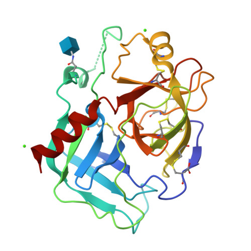
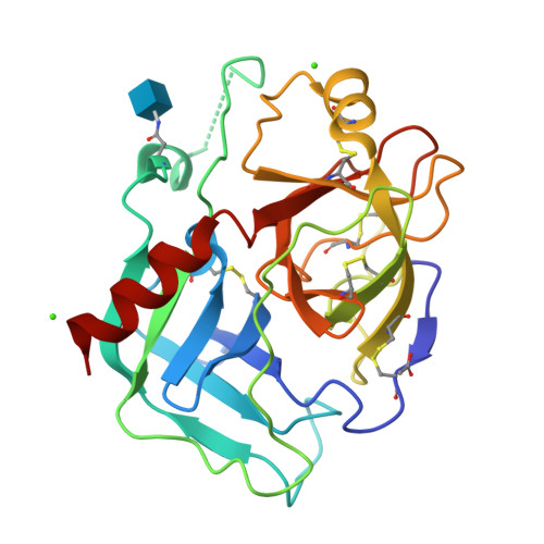
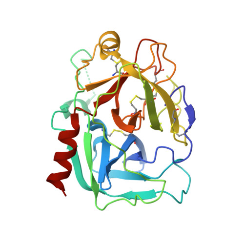
STRUCTURE OF MATURE HUMAN TISSUE KALLIKREIN (HUMAN KALLIKREIN 1 OR KLK1) AT 1.70 ANGSTROM RESOLUTION WITH VACANT ACTIVE SITE


Experimental Data Snapshot

  • Method: X-RAY DIFFRACTION
  • Resolution: 1.70 Å
  • R-Value Free: 0.202 
  • R-Value Work: 0.172 
  • R-Value Observed: 0.172 

wwPDB Validation   3D Report Full Report


Ligand Structure Quality Assessment 

Created with Raphaël 2.3.0Worse 01 BetterLigand structure goodness of fit to experimental dataBest fitted NAGClick on this verticalbar to view details

This is version 1.5 of the entry. See complete history


Literature

1.70 A X-ray structure of human apo kallikrein 1: Structural changes upon peptide inhibitor/substrate binding

Laxmikanthan, G.Blaber, S.I.Bernett, M.J.Scarisbrick, I.A.Juliano, M.A.Blaber, M.

(2005) Proteins 58: 802-814

  • DOI: https://doi.org/10.1002/prot.20368
  • Primary Citation of Related Structures:  
    1SPJ

  • PubMed Abstract: 

    Human kallikreins are serine proteases that comprise a recently identified large and closely related 15-member family. The kallikreins include both regulatory- and degradative-type proteases, impacting a variety of physiological processes including regulation of blood pressure, neuronal health, and the inflammatory response. While the function of the majority of the kallikreins remains to be elucidated, two members are useful biomarkers for prostate cancer and several others are potentially useful biomarkers for breast cancer, Alzheimer's, and Parkinson's disease. Human tissue kallikrein (human K1) is the best functionally characterized member of this family, and is known to play an important role in blood pressure regulation. As part of this function, human K1 exhibits unique dual-substrate specificity in hydrolyzing low molecular weight kininogen between both Arg-Ser and Met-Lys sequences. We report the X-ray crystal structure of mature, active recombinant human apo K1 at 1.70 A resolution. The active site exhibits structural features intermediate between that of apo and pro forms of known kallikrein structures. The S2 to S2' pockets demonstrate a variety of conformational changes in comparison to the porcine homolog of K1 in complex with peptide inhibitors, including the displacement of an extensive solvent network. These results indicate that the binding of a peptide substrate contributes to a structural rearrangement of the active-site Ser 195 resulting in a catalytically competent juxtaposition with the active-site His 57. The solvent networks within the S1 and S1' pockets suggest how the Arg-Ser and Met-Lys dual substrate specificity of human K1 is accommodated.


  • Organizational Affiliation

    Institute of Molecular Biophysics Florida State University, Tallahassee, Florida 32306-3015, USA.


Macromolecules
Find similar proteins by:  (by identity cutoff)  |  3D Structure
Entity ID: 1
MoleculeChains Sequence LengthOrganismDetailsImage
Kallikrein 1238Homo sapiensMutation(s): 2 
Gene Names: KLK1
EC: 3.4.21.35
UniProt & NIH Common Fund Data Resources
Find proteins for P06870 (Homo sapiens)
Explore P06870 
Go to UniProtKB:  P06870
PHAROS:  P06870
GTEx:  ENSG00000167748 
Entity Groups  
Sequence Clusters30% Identity50% Identity70% Identity90% Identity95% Identity100% Identity
UniProt GroupP06870
Glycosylation
Glycosylation Sites: 1Go to GlyGen: P06870-1
Sequence Annotations
Expand
  • Reference Sequence
Experimental Data & Validation

Experimental Data

  • Method: X-RAY DIFFRACTION
  • Resolution: 1.70 Å
  • R-Value Free: 0.202 
  • R-Value Work: 0.172 
  • R-Value Observed: 0.172 
  • Space Group: P 21 21 21
Unit Cell:
Length ( Å )Angle ( ˚ )
a = 44.719α = 90
b = 76.404β = 90
c = 76.626γ = 90
Software Package:
Software NamePurpose
DENZOdata reduction
SCALEPACKdata scaling
CNSrefinement
CNSphasing

Structure Validation

View Full Validation Report


Ligand Structure Quality Assessment 

Created with Raphaël 2.3.0Worse 01 BetterLigand structure goodness of fit to experimental dataBest fitted NAGClick on this verticalbar to view details

Entry History 

Deposition Data

Revision History  (Full details and data files)

  • Version 1.0: 2005-01-25
    Type: Initial release
  • Version 1.1: 2008-04-29
    Changes: Version format compliance
  • Version 1.2: 2011-07-13
    Changes: Non-polymer description, Version format compliance
  • Version 1.3: 2020-07-29
    Type: Remediation
    Reason: Carbohydrate remediation
    Changes: Data collection, Derived calculations, Structure summary
  • Version 1.4: 2021-10-27
    Changes: Database references, Structure summary
  • Version 1.5: 2024-10-30
    Changes: Data collection, Structure summary