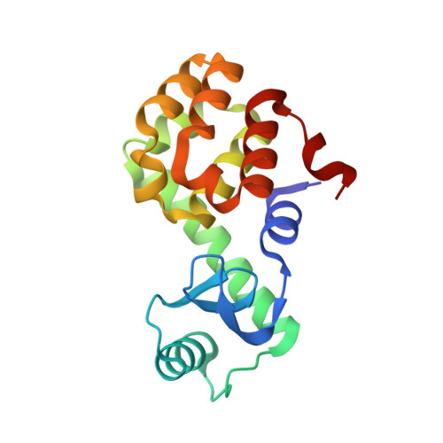
1SSW

Crystal structure of phage T4 lysozyme mutant Y24A/Y25A/T26A/I27A/C54T/C97A


Experimental Data Snapshot

  • Method: X-RAY DIFFRACTION
  • Resolution: 2.13 Å
  • R-Value Free: 0.215 
  • R-Value Work: 0.187 
  • R-Value Observed: 0.187 

wwPDB Validation   3D Report Full Report


This is version 1.6 of the entry. See complete history


Literature

Alanine-scanning mutagenesis of the beta-sheet region of phage T4 lysozyme suggests that tertiary context has a dominant effect on beta-sheet formation

He, M.M.Wood, Z.A.Baase, W.A.Xiao, H.Matthews, B.W.

(2004) Protein Sci 13: 2716-2724

  • DOI: https://doi.org/10.1110/ps.04875504
  • Primary Citation of Related Structures:  
    1SSW, 1SSY, 1T8F, 1T8G

  • PubMed Abstract: 

    In general, alpha-helical conformations in proteins depend in large part on the amino acid residues within the helix and their proximal interactions. For example, an alanine residue has a high propensity to adopt an alpha-helical conformation, whereas that of a glycine residue is low. The sequence preferences for beta-sheet formation are less obvious. To identify the factors that influence beta-sheet conformation, a series of scanning polyalanine mutations were made within the strands and associated turns of the beta-sheet region in T4 lysozyme. For each construct the stability of the folded protein was reduced substantially, consistent with removal of native packing interactions. However, the crystal structures showed that each of the mutants retained the beta-sheet conformation. These results suggest that the structure of the beta-sheet region of T4 lysozyme is maintained to a substantial extent by tertiary interactions with the surrounding parts of the protein. Such tertiary interactions may be important in determining the structures of beta-sheets in general.


  • Organizational Affiliation

    Institute of Molecular Biology, Howard Hughes Medical Institute and Department of Physics, 1229 University of Oregon, Eugene, OR 97403-1229, USA.


Macromolecules
Find similar proteins by:  (by identity cutoff)  |  3D Structure
Entity ID: 1
MoleculeChains Sequence LengthOrganismDetailsImage
Lysozyme164Tequatrovirus T4Mutation(s): 6 
EC: 3.2.1.17
UniProt
Find proteins for P00720 (Enterobacteria phage T4)
Explore P00720 
Go to UniProtKB:  P00720
Entity Groups  
Sequence Clusters30% Identity50% Identity70% Identity90% Identity95% Identity100% Identity
UniProt GroupP00720
Sequence Annotations
Expand
  • Reference Sequence
Small Molecules
Ligands 1 Unique
IDChains Name / Formula / InChI Key2D Diagram3D Interactions
BME
Query on BME

Download Ideal Coordinates CCD File 
B [auth A]BETA-MERCAPTOETHANOL
C2 H6 O S
DGVVWUTYPXICAM-UHFFFAOYSA-N
Experimental Data & Validation

Experimental Data

  • Method: X-RAY DIFFRACTION
  • Resolution: 2.13 Å
  • R-Value Free: 0.215 
  • R-Value Work: 0.187 
  • R-Value Observed: 0.187 
  • Space Group: P 32 2 1
Unit Cell:
Length ( Å )Angle ( ˚ )
a = 60.841α = 90
b = 60.841β = 90
c = 96.131γ = 120
Software Package:
Software NamePurpose
CNSrefinement
MOSFLMdata reduction
CCP4data scaling
AMoREphasing
REFMACrefinement

Structure Validation

View Full Validation Report


Entry History 

Deposition Data

Revision History  (Full details and data files)

  • Version 1.0: 2004-10-19
    Type: Initial release
  • Version 1.1: 2008-04-29
    Changes: Version format compliance
  • Version 1.2: 2011-07-13
    Changes: Version format compliance
  • Version 1.3: 2012-04-11
    Changes: Version format compliance
  • Version 1.4: 2019-07-24
    Changes: Data collection, Refinement description
  • Version 1.5: 2021-10-27
    Changes: Database references, Derived calculations
  • Version 1.6: 2024-02-14
    Changes: Data collection