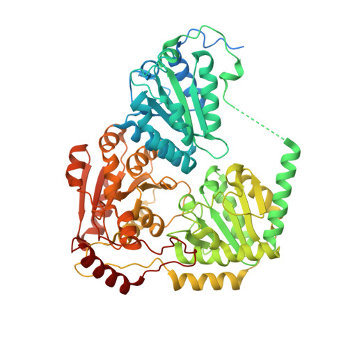
1T9C

Crystal Structure Of Yeast Acetohydroxyacid Synthase In Complex With A Sulfonylurea Herbicide, Sulfometuron methyl


Experimental Data Snapshot

  • Method: X-RAY DIFFRACTION
  • Resolution: 2.34 Å
  • R-Value Free: 0.191 
  • R-Value Work: 0.162 
  • R-Value Observed: 0.164 

Starting Model: experimental
View more details

wwPDB Validation   3D Report Full Report


Ligand Structure Quality Assessment 


This is version 1.4 of the entry. See complete history


Literature

Elucidating the specificity of binding of sulfonylurea herbicides to acetohydroxyacid synthase.

McCourt, J.A.Pang, S.S.Guddat, L.W.Duggleby, R.G.

(2005) Biochemistry 44: 2330-2338

  • DOI: https://doi.org/10.1021/bi047980a
  • Primary Citation of Related Structures:  
    1T9A, 1T9B, 1T9C, 1T9D

  • PubMed Abstract: 

    Acetohydroxyacid synthase (AHAS, EC 2.2.1.6) is the target for the sulfonylurea herbicides, which act as potent inhibitors of the enzyme. Chlorsulfuron (marketed as Glean) and sulfometuron methyl (marketed as Oust) are two commercially important members of this family of herbicides. Here we report crystal structures of yeast AHAS in complex with chlorsulfuron (at a resolution of 2.19 A), sulfometuron methyl (2.34 A), and two other sulfonylureas, metsulfuron methyl (2.29 A) and tribenuron methyl (2.58 A). The structures observed suggest why these inhibitors have different potencies and provide clues about the differential effects of mutations in the active site tunnel on various inhibitors. In all of the structures, the thiamin diphosphate cofactor is fragmented, possibly as the result of inhibitor binding. In addition to thiamin diphosphate, AHAS requires FAD for activity. Recently, it has been reported that reduction of FAD can occur as a minor side reaction due to reaction with the carbanion/enamine of the hydroxyethyl-ThDP intermediate that is formed midway through the catalytic cycle. Here we report that the isoalloxazine ring has a bent conformation that would account for its ability to accept electrons from the hydroxyethyl intermediate. Most sequence and mutation data suggest that yeast AHAS is a high-quality model for the plant enzyme.


  • Organizational Affiliation

    Department of Biochemistry and Molecular Biology, The University of Queensland, Brisbane, Queensland 4072, Australia.


Macromolecules
Find similar proteins by:  (by identity cutoff)  |  3D Structure
Entity ID: 1
MoleculeChains Sequence LengthOrganismDetailsImage
Acetolactate synthase, mitochondrial
A, B
677Saccharomyces cerevisiaeMutation(s): 0 
Gene Names: ILV2SMR1YMR108WYM9718.07
EC: 2.2.1.6
UniProt
Find proteins for P07342 (Saccharomyces cerevisiae (strain ATCC 204508 / S288c))
Explore P07342 
Go to UniProtKB:  P07342
Entity Groups  
Sequence Clusters30% Identity50% Identity70% Identity90% Identity95% Identity100% Identity
UniProt GroupP07342
Sequence Annotations
Expand
  • Reference Sequence
Small Molecules
Ligands 6 Unique
IDChains Name / Formula / InChI Key2D Diagram3D Interactions
FAD
Query on FAD

Download Ideal Coordinates CCD File 
F [auth A],
L [auth B]
FLAVIN-ADENINE DINUCLEOTIDE
C27 H33 N9 O15 P2
VWWQXMAJTJZDQX-UYBVJOGSSA-N
1SM
Query on 1SM

Download Ideal Coordinates CCD File 
E [auth A],
K [auth B]
METHYL 2-[({[(4,6-DIMETHYLPYRIMIDIN-2-YL)AMINO]CARBONYL}AMINO)SULFONYL]BENZOATE
C15 H16 N4 O5 S
ZDXMLEQEMNLCQG-UHFFFAOYSA-N
P23
Query on P23

Download Ideal Coordinates CCD File 
J [auth B]PROPYL TRIHYDROGEN DIPHOSPHATE
C3 H10 O7 P2
JCWMAIAFQQTSAX-UHFFFAOYSA-N
P22
Query on P22

Download Ideal Coordinates CCD File 
G [auth A]ETHYL DIHYDROGEN DIPHOSPHATE
C2 H8 O7 P2
OJDJHGIXNZPZFD-UHFFFAOYSA-N
K
Query on K

Download Ideal Coordinates CCD File 
C [auth A],
H [auth B]
POTASSIUM ION
K
NPYPAHLBTDXSSS-UHFFFAOYSA-N
MG
Query on MG

Download Ideal Coordinates CCD File 
D [auth A],
I [auth B]
MAGNESIUM ION
Mg
JLVVSXFLKOJNIY-UHFFFAOYSA-N
Binding Affinity Annotations 
IDSourceBinding Affinity
1SM BindingDB:  1T9C Ki: 51 (nM) from 1 assay(s)
Experimental Data & Validation

Experimental Data

  • Method: X-RAY DIFFRACTION
  • Resolution: 2.34 Å
  • R-Value Free: 0.191 
  • R-Value Work: 0.162 
  • R-Value Observed: 0.164 
  • Space Group: P 4 2 2
Unit Cell:
Length ( Å )Angle ( ˚ )
a = 154.486α = 90
b = 154.486β = 90
c = 178.796γ = 90
Software Package:
Software NamePurpose
Adxvdata processing
SCALEPACKdata scaling
AMoREphasing
CNSrefinement

Structure Validation

View Full Validation Report


Ligand Structure Quality Assessment 


Entry History 

Deposition Data

Revision History  (Full details and data files)

  • Version 1.0: 2004-12-21
    Type: Initial release
  • Version 1.1: 2008-04-30
    Changes: Version format compliance
  • Version 1.2: 2011-07-13
    Changes: Version format compliance
  • Version 1.3: 2017-10-11
    Changes: Refinement description
  • Version 1.4: 2023-08-23
    Changes: Data collection, Database references, Derived calculations, Refinement description