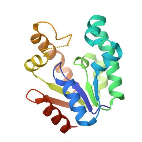
Crystallographic studies of shikimate binding and induced conformational changes in Mycobacterium tuberculosis shikimate kinase.
Dhaliwal, B., Nichols, C.E., Ren, J., Lockyer, M., Charles, I., Hawkins, A.R., Stammers, D.K.(2004) FEBS Lett 574: 49-54
- PubMed: 15358538
- DOI: https://doi.org/10.1016/j.febslet.2004.08.005
- Primary Citation of Related Structures:
1U8A - PubMed Abstract:
The X-ray crystal structure of Mycobacterium tuberculosis shikimate kinase (SK) with bound shikimate and adenosine diphosphate (ADP) has been determined to a resolution of 2.15 A. The binding of shikimate in a shikimate kinase crystal structure has not previously been reported. The substrate binds in a pocket lined with hydrophobic residues and interacts with several highly conserved charged residues including Asp34, Arg58, Glu61 and Arg136 which project into the cavity. Comparisons of our ternary SK-ADP-shikimate complex with an earlier binary SK-ADP complex show that conformational changes occur on shikimate binding with the substrate-binding domain rotating by 10 degrees. Detailed knowledge of shikimate binding is an important step in the design of inhibitors of SK, which have potential as novel anti-tuberculosis agents.
- Division of Structural Biology, The Wellcome Trust Centre for Human Genetics, University of Oxford, Roosevelt Drive, Oxford OX3 7BN, UK.
Organizational Affiliation:


