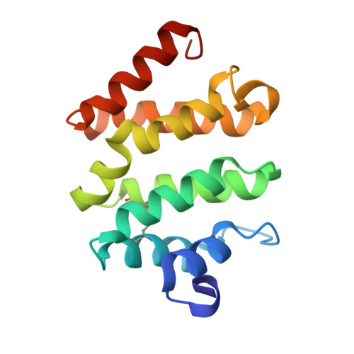
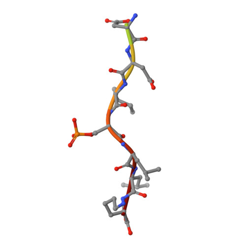
1UJK

VHS domain of human GGA1 complexed with C-terminal phosphopeptide from BACE


Experimental Data Snapshot

  • Method: X-RAY DIFFRACTION
  • Resolution: 1.90 Å
  • R-Value Free: 0.245 
  • R-Value Work: 0.213 
  • R-Value Observed: 0.215 

Starting Model: experimental
View more details

wwPDB Validation   3D Report Full Report


This is version 1.4 of the entry. See complete history


Literature

Insights into the Phosphoregulation of beta-Secretase Sorting Signal by the VHS Domain of GGA1

Shiba, T.Kametaka, S.Kawasaki, M.Shibata, M.Waguri, S.Uchiyama, Y.Wakatsuki, S.

(2004) Traffic 5: 437-448

  • DOI: https://doi.org/10.1111/j.1600-0854.2004.00188.x
  • Primary Citation of Related Structures:  
    1UJJ, 1UJK

  • PubMed Abstract: 

    BACE (beta-site amyloid precursor protein cleaving enzyme, beta-secretase) is a type-I membrane protein which functions as an aspartic protease in the production of beta-amyloid peptide, a causative agent of Alzheimer's disease. Its cytoplasmic tail has a characteristic acidic-cluster dileucine motif recognized by the VHS domain of adaptor proteins, GGAs (Golgi-localizing, gamma-adaptin ear homology domain, ARF-interacting). Here we show that BACE is colocalized with GGAs in the trans-Golgi network and peripheral structures, and phosphorylation of a serine residue in the cytoplasmic tail enhances interaction with the VHS domain of GGA1 by about threefold. The X-ray crystal structure of the complex between the GGA1-VHS domain and the BACE C-terminal peptide illustrates a similar recognition mechanism as mannose 6-phosphate receptors except that a glutamine residue closes in to fill the gap created by the shorter BACE peptide. The serine and lysine of the BACE peptide point their side chains towards the solvent. However, phosphorylation of the serine affects the lysine side chain and the peptide backbone, resulting in one additional hydrogen bond and a stronger electrostatic interaction with the VHS domain, hence the reversible increase in affinity.


  • Organizational Affiliation

    Structural Biology Research Center, Photon Factory, Institute of Materials Structure Science, High Energy Accelerator Research Organization (KEK), Tsukuba, Ibaraki 305-0801, Japan.


Macromolecules
Find similar proteins by:  (by identity cutoff)  |  3D Structure
Entity ID: 1
MoleculeChains Sequence LengthOrganismDetailsImage
ADP-ribosylation factor binding protein GGA1
A, B
147Homo sapiensMutation(s): 0 
UniProt & NIH Common Fund Data Resources
Find proteins for Q9UJY5 (Homo sapiens)
Explore Q9UJY5 
Go to UniProtKB:  Q9UJY5
PHAROS:  Q9UJY5
GTEx:  ENSG00000100083 
Entity Groups  
Sequence Clusters30% Identity50% Identity70% Identity90% Identity95% Identity100% Identity
UniProt GroupQ9UJY5
Sequence Annotations
Expand
  • Reference Sequence

Find similar proteins by:  Sequence   |   3D Structure  

Entity ID: 2
MoleculeChains Sequence LengthOrganismDetailsImage
C-terminal peptide from Beta-secretase
C, D
12N/AMutation(s): 1 
EC: 3.4.23 (PDB Primary Data), 3.4.23.46 (UniProt)
UniProt & NIH Common Fund Data Resources
Find proteins for P56817 (Homo sapiens)
Explore P56817 
Go to UniProtKB:  P56817
PHAROS:  P56817
GTEx:  ENSG00000186318 
Entity Groups  
Sequence Clusters30% Identity50% Identity70% Identity90% Identity95% Identity100% Identity
UniProt GroupP56817
Sequence Annotations
Expand
  • Reference Sequence
Experimental Data & Validation

Experimental Data

  • Method: X-RAY DIFFRACTION
  • Resolution: 1.90 Å
  • R-Value Free: 0.245 
  • R-Value Work: 0.213 
  • R-Value Observed: 0.215 
  • Space Group: P 21 21 21
Unit Cell:
Length ( Å )Angle ( ˚ )
a = 48.545α = 90
b = 72.308β = 90
c = 103.13γ = 90
Software Package:
Software NamePurpose
HKL-2000data collection
SCALAdata scaling
MOLREPphasing
CNSrefinement
HKL-2000data reduction
CCP4data scaling

Structure Validation

View Full Validation Report


Entry History 

Deposition Data

Revision History  (Full details and data files)

  • Version 1.0: 2004-05-11
    Type: Initial release
  • Version 1.1: 2008-04-27
    Changes: Version format compliance
  • Version 1.2: 2011-07-13
    Changes: Version format compliance
  • Version 1.3: 2023-10-25
    Changes: Data collection, Database references, Derived calculations, Refinement description
  • Version 1.4: 2024-10-30
    Changes: Structure summary