1UR0 | pdb_00001ur0

The structure of endo-beta-1,4-galactanase from Bacillus licheniformis in complex with two oligosaccharide products.


Experimental Data Snapshot

  • Method: X-RAY DIFFRACTION
  • Resolution: 2.50 Å
  • R-Value Free: 
    0.244 (Depositor) 
  • R-Value Work: 
    0.204 (Depositor) 
  • R-Value Observed: 
    0.204 (Depositor) 

wwPDB Validation 3D Report Full Report

Validation slider image for 1UR0

This is version 2.1 of the entry. See complete history

Literature

The Structure of Endo-Beta-1,4-Galactanase from Bacillus Licheniformis in Complex with Two Oligosaccharide Products

Ryttersgaard, C.Le Nours, J.Lo Leggio, L.Jorgensen, C.T.Christensen, L.L.H.Bjornvad, M.Larsen, S.

(2004) J Mol Biology 341: 107

  • DOI: https://doi.org/10.1016/j.jmb.2004.05.017
  • Primary Citation Related Structures: 
    1R8L, 1UR0, 1UR4

  • PubMed Abstract: 

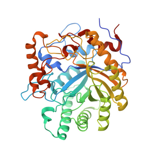
    The beta-1,4-galactanase from Bacillus licheniformis (BLGAL) is a plant cell-wall-degrading enzyme involved in the hydrolysis of beta-1,4-galactan in the hairy regions of pectin. The crystal structure of BLGAL was determined by molecular replacement both alone and in complex with the products galactobiose and galactotriose, catching a first crystallographic glimpse of fragments of beta-1,4-galactan. As expected for an enzyme belonging to GH-53, the BLGAL structure reveals a (betaalpha)(8)-barrel architecture. However, BLGAL betaalpha-loops 2, 7 and 8 are long in contrast to the corresponding loops in structures of fungal galactanases determined previously. The structure of BLGAL additionally shows a calcium ion linking the long betaalpha-loops 7 and 8, which replaces a disulphide bridge in the fungal galactanases. Compared to the substrate-binding subsites predicted for Aspergillus aculeatus galactanase (AAGAL), two additional subsites for substrate binding are found in BLGAL, -3 and -4. A comparison of the pattern of galactan and galactooligosaccharides degradation by AAGAL and BLGAL shows that, although both are most active on substrates with a high degree of polymerization, AAGAL can degrade galactotriose and galactotetraose efficiently, whereas BLGAL prefers longer oligosaccharides and cannot hydrolyze galactotriose to any appreciable extent. This difference in substrate preference can be explained structurally by the presence of the extra subsites -3 and -4 in BLGAL.


  • Organizational Affiliation
    • Centre for Crystallographic Studies, Department of Chemistry, University of Copenhagen, Universitetsparken 5, DK-2100 Copenhagen, Denmark.

Macromolecule Content 

  • Total Structure Weight: 88.95 kDa 
  • Atom Count: 6,233 
  • Modeled Residue Count: 775 
  • Deposited Residue Count: 798 
  • Unique protein chains: 1

Macromolecules

Find similar proteins by:|  3D Structure
Entity ID: 1
MoleculeChains  Sequence LengthOrganismDetailsImage
GALACTANASE
A, B
399Bacillus licheniformisMutation(s): 0 
EC: 3.2.1.89 (PDB Primary Data), 3.2.1 (UniProt)
UniProt
Find proteins for Q65CX5 (Bacillus licheniformis (strain ATCC 14580 / DSM 13 / JCM 2505 / CCUG 7422 / NBRC 12200 / NCIMB 9375 / NCTC 10341 / NRRL NRS-1264 / Gibson 46))
Explore Q65CX5 
Go to UniProtKB:  Q65CX5
Entity Groups
Sequence Clusters30% Identity50% Identity70% Identity90% Identity95% Identity100% Identity
UniProt GroupQ65CX5
Sequence Annotations
Expand
Reference Sequence

Oligosaccharides

Help  
Entity ID: 2
MoleculeChains Length2D Diagram GlycosylationD Interactions
beta-D-galactopyranose-(1-4)-beta-D-galactopyranose-(1-4)-beta-D-galactopyranose
C, D
3N/A
Glycosylation Resources
GlyTouCan: G06996VN
GlyCosmos: G06996VN
GlyGen: G06996VN

Biologically Interesting Molecules (External Reference) 

1 Unique

Experimental Data & Validation

Experimental Data

  • Method: X-RAY DIFFRACTION
  • Resolution: 2.50 Å
  • R-Value Free:  0.244 (Depositor) 
  • R-Value Work:  0.204 (Depositor) 
  • R-Value Observed: 0.204 (Depositor) 
Space Group: P 1 21 1
Unit Cell:
Length ( Å )Angle ( ˚ )
a = 50.57α = 90
b = 80.14β = 99.39
c = 103.22γ = 90
Software Package:
Software NamePurpose
CNSrefinement
DENZOdata reduction
SCALEPACKdata scaling

Structure Validation

View Full Validation Report


Entry History 

Deposition Data

Revision History  (Full details and data files)

  • Version 1.0: 2004-10-20
    Type: Initial release
  • Version 1.1: 2011-05-08
    Changes: Version format compliance
  • Version 1.2: 2011-07-13
    Changes: Version format compliance
  • Version 1.3: 2019-07-24
    Changes: Data collection
  • Version 2.0: 2020-07-29
    Type: Remediation
    Reason: Carbohydrate remediation
    Changes: Atomic model, Data collection, Derived calculations, Non-polymer description, Structure summary
  • Version 2.1: 2024-05-08
    Changes: Data collection, Database references, Structure summary