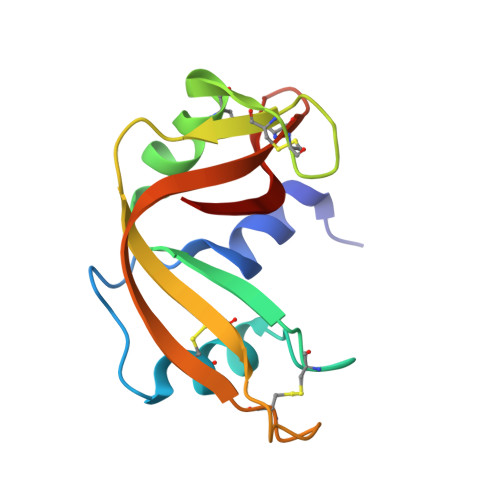
1W4Q

Binding of Nonnatural 3'-Nucleotides to Ribonuclease A


Experimental Data Snapshot

  • Method: X-RAY DIFFRACTION
  • Resolution: 1.68 Å
  • R-Value Free: 0.241 
  • R-Value Work: 0.209 
  • R-Value Observed: 0.209 

Starting Model: experimental
View more details

wwPDB Validation   3D Report Full Report


Ligand Structure Quality Assessment 


This is version 1.3 of the entry. See complete history


Literature

Binding of Non-Natural 3'-Nucleotides to Ribonuclease A

Jenkins, C.L.Thiyagarajan, N.Sweeney, R.Y.Guy, M.P.Kelemen, B.R.Acharya, K.R.Raines, R.T.

(2005) FEBS J 272: 744

  • DOI: https://doi.org/10.1111/j.1742-4658.2004.04511.x
  • Primary Citation of Related Structures:  
    1W4O, 1W4P, 1W4Q

  • PubMed Abstract: 

    2'-Fluoro-2'-deoxyuridine 3'-phosphate (dU(F)MP) and arabinouridine 3'-phosphate (araUMP) have non-natural furanose rings. dU(F)MP and araUMP were prepared by chemical synthesis and found to have three- to sevenfold higher affinity than uridine 3'-phosphate (3'-UMP) or 2'-deoxyuridine 3'-phosphate (dUMP) for ribonuclease A (RNase A). These differences probably arise (in part) from the phosphoryl groups of 3'-UMP, dU(F)MP, and araUMP (pK(a) = 5.9) being more anionic than that of dUMP (pK(a) = 6.3). The three-dimensional structures of the crystalline complexes of RNase A with dUMP, dU(F)MP and araUMP were determined at < 1.7 A resolution by X-ray diffraction analysis. In these three structures, the uracil nucleobases and phosphoryl groups bind to the enzyme in a nearly identical position. Unlike 3'-UMP and dU(F)MP, dUMP and araUMP bind with their furanose rings in the preferred pucker. In the RNase A.araUMP complex, the 2'-hydroxyl group is exposed to the solvent. All four 3'-nucleotides bind more tightly to wild-type RNase A than to its T45G variant, which lacks the residue that interacts most closely with the uracil nucleobase. These findings illuminate in atomic detail the interaction of RNase A and 3'-nucleotides, and indicate that non-natural furanose rings can serve as the basis for more potent inhibitors of catalysis by RNase A.


  • Organizational Affiliation

    Department of Chemistry, University of Wisconsin-Madison, WI, USA.


Macromolecules
Find similar proteins by:  (by identity cutoff)  |  3D Structure
Entity ID: 1
MoleculeChains Sequence LengthOrganismDetailsImage
PANCREATIC RIBONUCLEASE A
A, B
124Bos taurusMutation(s): 0 
EC: 3.1.27.5 (PDB Primary Data), 4.6.1.18 (UniProt)
UniProt
Find proteins for P61823 (Bos taurus)
Explore P61823 
Go to UniProtKB:  P61823
Entity Groups  
Sequence Clusters30% Identity50% Identity70% Identity90% Identity95% Identity100% Identity
UniProt GroupP61823
Sequence Annotations
Expand
  • Reference Sequence
Small Molecules
Binding Affinity Annotations 
IDSourceBinding Affinity
UMF PDBBind:  1W4Q Ki: 5500 (nM) from 1 assay(s)
Experimental Data & Validation

Experimental Data

  • Method: X-RAY DIFFRACTION
  • Resolution: 1.68 Å
  • R-Value Free: 0.241 
  • R-Value Work: 0.209 
  • R-Value Observed: 0.209 
  • Space Group: C 1 2 1
Unit Cell:
Length ( Å )Angle ( ˚ )
a = 100.101α = 90
b = 32.925β = 90.39
c = 72.787γ = 90
Software Package:
Software NamePurpose
CNSrefinement
HKL-2000data reduction
HKL-2000data scaling
AMoREphasing

Structure Validation

View Full Validation Report


Ligand Structure Quality Assessment 


Entry History 

Deposition Data

Revision History  (Full details and data files)

  • Version 1.0: 2005-02-02
    Type: Initial release
  • Version 1.1: 2011-05-08
    Changes: Version format compliance
  • Version 1.2: 2011-07-13
    Changes: Version format compliance
  • Version 1.3: 2023-12-13
    Changes: Data collection, Database references, Other, Refinement description