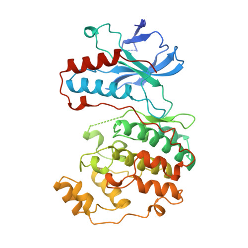
1WFC

STRUCTURE OF APO, UNPHOSPHORYLATED, P38 MITOGEN ACTIVATED PROTEIN KINASE P38 (P38 MAP KINASE) THE MAMMALIAN HOMOLOGUE OF THE YEAST HOG1 PROTEIN


Experimental Data Snapshot

  • Method: X-RAY DIFFRACTION
  • Resolution: 2.30 Å
  • R-Value Free: 0.297 
  • R-Value Work: 0.213 
  • R-Value Observed: 0.213 

wwPDB Validation   3D Report Full Report


This is version 1.3 of the entry. See complete history


Literature

Crystal structure of p38 mitogen-activated protein kinase.

Wilson, K.P.Fitzgibbon, M.J.Caron, P.R.Griffith, J.P.Chen, W.McCaffrey, P.G.Chambers, S.P.Su, M.S.

(1996) J Biol Chem 271: 27696-27700

  • DOI: https://doi.org/10.1074/jbc.271.44.27696
  • Primary Citation of Related Structures:  
    1WFC

  • PubMed Abstract: 

    p38 mitogen-activated protein kinase is activated by environmental stress and cytokines and plays a role in transcriptional regulation and inflammatory responses. The crystal structure of the apo, unphosphorylated form of p38 kinase has been solved at 2.3 A resolution. The fold and topology of p38 is similar to ERK2 (Zhang, F., Strand, A., Robbins, D., Cobb, M. H., and Goldsmith, E. J. (1994) Nature 367, 704-711). The relative orientation of the two domains of p38 kinase is different from that observed in the active form of cAMP-dependent protein kinase. The twist results in a misalignment of the active site of p38, suggesting that the orientation of the domains would have to change before catalysis could proceed. The residues that are phosphorylated upon activation of p38 are located on a surface loop that occupies the peptide binding channel. Occlusion of the active site by the loop, and misalignment of catalytic residues, may account for the low enzymatic activity of unphosphorylated p38 kinase.


  • Organizational Affiliation

    Vertex Pharmaceuticals Incorporated, Cambridge, Massachusetts 02139-4211, USA.


Macromolecules
Find similar proteins by:  (by identity cutoff)  |  3D Structure
Entity ID: 1
MoleculeChains Sequence LengthOrganismDetailsImage
MITOGEN-ACTIVATED PROTEIN KINASE P38366Homo sapiensMutation(s): 0 
EC: 2.7.1 (PDB Primary Data), 2.7.11.24 (UniProt)
UniProt & NIH Common Fund Data Resources
Find proteins for Q16539 (Homo sapiens)
Explore Q16539 
Go to UniProtKB:  Q16539
PHAROS:  Q16539
GTEx:  ENSG00000112062 
Entity Groups  
Sequence Clusters30% Identity50% Identity70% Identity90% Identity95% Identity100% Identity
UniProt GroupQ16539
Sequence Annotations
Expand
  • Reference Sequence
Experimental Data & Validation

Experimental Data

  • Method: X-RAY DIFFRACTION
  • Resolution: 2.30 Å
  • R-Value Free: 0.297 
  • R-Value Work: 0.213 
  • R-Value Observed: 0.213 
  • Space Group: P 21 21 21
Unit Cell:
Length ( Å )Angle ( ˚ )
a = 45.193α = 90
b = 86.25β = 90
c = 123.988γ = 90
Software Package:
Software NamePurpose
X-PLORmodel building
X-PLORrefinement
bioteXdata reduction
bioteXdata scaling
X-PLORphasing

Structure Validation

View Full Validation Report


Entry History 

Deposition Data

Revision History  (Full details and data files)

  • Version 1.0: 1997-09-19
    Type: Initial release
  • Version 1.1: 2008-03-24
    Changes: Version format compliance
  • Version 1.2: 2011-07-13
    Changes: Version format compliance
  • Version 1.3: 2024-02-14
    Changes: Data collection, Database references, Other, Refinement description