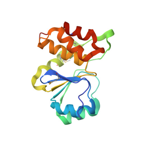
1XRI

X-ray structure of a putative phosphoprotein phosphatase from Arabidopsis thaliana gene AT1G05000


Experimental Data Snapshot

  • Method: X-RAY DIFFRACTION
  • Resolution: 3.30 Å
  • R-Value Free: 0.249 
  • R-Value Work: 0.204 

wwPDB Validation   3D Report Full Report


This is version 1.5 of the entry. See complete history

Re-refinement Note

A newer entry is available that reflects an alternative modeling of the original data: 2Q47


Literature

Structural and functional characterization of a novel phosphatase from the Arabidopsis thaliana gene locus At1g05000.

Aceti, D.J.Bitto, E.Yakunin, A.F.Proudfoot, M.Bingman, C.A.Frederick, R.O.Sreenath, H.K.Vojtik, F.C.Wrobel, R.L.Fox, B.G.Markley, J.L.Phillips Jr., G.N.

(2008) Proteins 73: 241-253

  • DOI: https://doi.org/10.1002/prot.22041
  • Primary Citation of Related Structures:  
    1XRI

  • PubMed Abstract: 

    The crystal structure of the protein product of the gene locus At1g05000, a hypothetical protein from A. thaliana, was determined by the multiple-wavelength anomalous diffraction method and was refined to an R factor of 20.4% (R(free) = 24.9%) at 3.3 A. The protein adopts the alpha/beta fold found in cysteine phosphatases, a superfamily of phosphatases that possess a catalytic cysteine and form a covalent thiol-phosphate intermediate during the catalytic cycle. In At1g05000, the analogous cysteine (Cys(150)) is located at the bottom of a positively-charged pocket formed by residues that include the conserved arginine (Arg(156)) of the signature active site motif, HCxxGxxRT. Of 74 model phosphatase substrates tested, purified recombinant At1g05000 showed highest activity toward polyphosphate (poly-P(12-13)) and deoxyribo- and ribonucleoside triphosphates, and less activity toward phosphoenolpyruvate, phosphotyrosine, phosphotyrosine-containing peptides, and phosphatidyl inositols. Divalent metal cations were not required for activity and had little effect on the reaction.


  • Organizational Affiliation

    Department of Biochemistry, The Center for Eukaryotic Structural Genomics, University of Wisconsin at Madison, Madison, Wisconsin 53706, USA.


Macromolecules
Find similar proteins by:  (by identity cutoff)  |  3D Structure
Entity ID: 1
MoleculeChains Sequence LengthOrganismDetailsImage
At1g05000
A, B
151Arabidopsis thalianaMutation(s): 0 
Gene Names: At1g05000
EC: 3.6.1.52
UniProt
Find proteins for Q9ZVN4 (Arabidopsis thaliana)
Explore Q9ZVN4 
Go to UniProtKB:  Q9ZVN4
Entity Groups  
Sequence Clusters30% Identity50% Identity70% Identity90% Identity95% Identity100% Identity
UniProt GroupQ9ZVN4
Sequence Annotations
Expand
  • Reference Sequence
Experimental Data & Validation

Experimental Data

  • Method: X-RAY DIFFRACTION
  • Resolution: 3.30 Å
  • R-Value Free: 0.249 
  • R-Value Work: 0.204 
  • Space Group: P 21 3
Unit Cell:
Length ( Å )Angle ( ˚ )
a = 124.483α = 90
b = 124.483β = 90
c = 124.483γ = 90
Software Package:
Software NamePurpose
DENZOdata reduction
SCALEPACKdata scaling
SOLVEphasing
RESOLVEphasing
CNSrefinement
PDB_EXTRACTdata extraction

Structure Validation

View Full Validation Report


Entry History 

Revision History  (Full details and data files)

  • Version 1.0: 2004-10-26
    Type: Initial release
  • Version 1.1: 2008-02-01
    Changes: Version format compliance
  • Version 1.2: 2011-07-13
    Changes: Derived calculations, Version format compliance
  • Version 1.3: 2013-02-06
    Changes: Database references
  • Version 1.4: 2017-10-11
    Changes: Refinement description
  • Version 1.5: 2024-10-16
    Changes: Data collection, Database references, Derived calculations, Structure summary