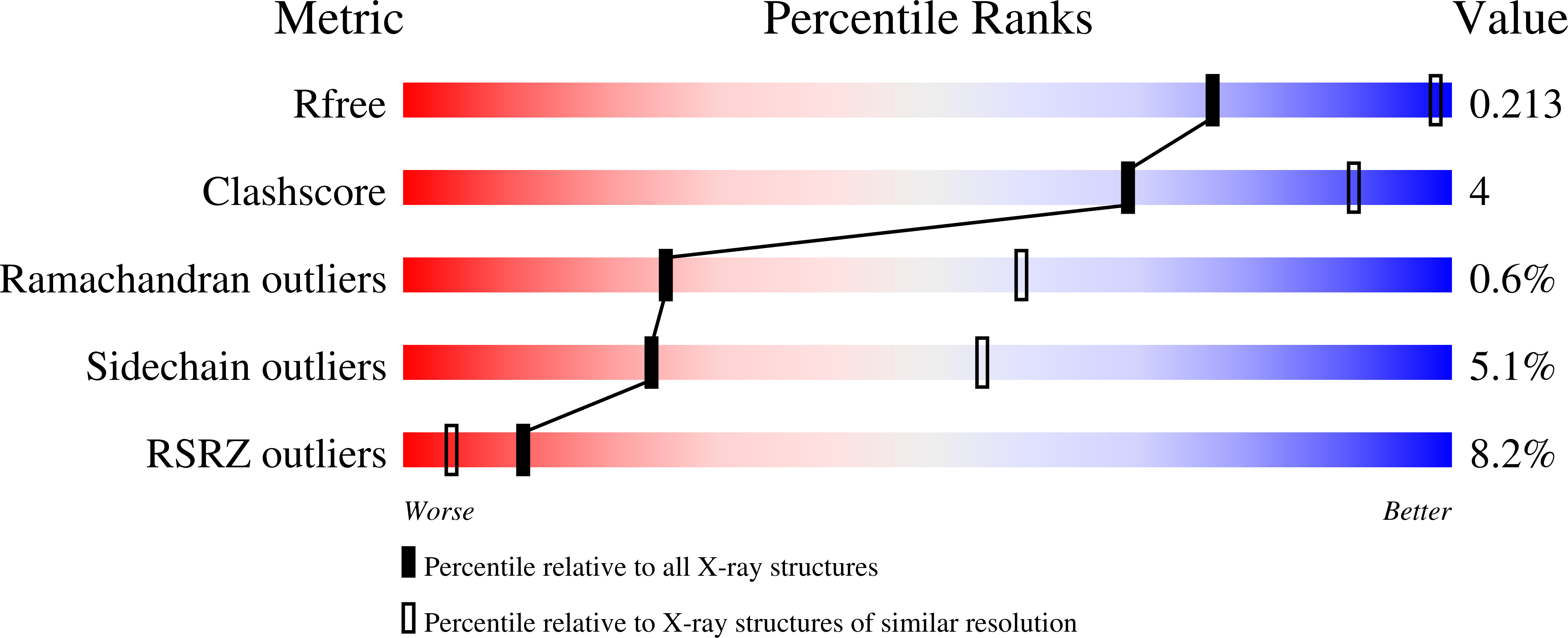
A double mutation of Escherichia coli2C-methyl-D-erythritol-2,4-cyclodiphosphate synthase disrupts six hydrogen bonds with, yet fails to prevent binding of, an isoprenoid diphosphate.
Sgraja, T., Kemp, L.E., Ramsden, N., Hunter, W.N.(2005) Acta Crystallogr Sect F Struct Biol Cryst Commun 61: 625-629
- PubMed: 16511114
- DOI: https://doi.org/10.1107/S1744309105018762
- Primary Citation of Related Structures:
1YQN - PubMed Abstract:
The essential enzyme 2C-methyl-D-erythritol-2,4-cyclodiphosphate (MECP) synthase, found in most eubacteria and the apicomplexan parasites, participates in isoprenoid-precursor biosynthesis and is a validated target for the development of broad-spectrum antimicrobial drugs. The structure and mechanism of the enzyme have been elucidated and the recent exciting finding that the enzyme actually binds diphosphate-containing isoprenoids at the interface formed by the three subunits that constitute the active protein suggests the possibility of feedback regulation of MECP synthase. To investigate such a possibility, a form of the enzyme was sought that did not bind these ligands but which would retain the quaternary structure necessary to create the active site. Two amino acids, Arg142 and Glu144, in Escherichia coli MECP synthase were identified as contributing to ligand binding. Glu144 interacts directly with Arg142 and positions the basic residue to form two hydrogen bonds with the terminal phosphate group of the isoprenoid diphosphate ligand. This association occurs at the trimer interface and three of these arginines interact with the ligand phosphate group. A dual mutation was designed (Arg142 to methionine and Glu144 to leucine) to disrupt the electrostatic attractions between the enzyme and the phosphate group to investigate whether an enzyme without isoprenoid diphosphate could be obtained. A low-resolution crystal structure of the mutated MECP synthase Met142/Leu144 revealed that geranyl diphosphate was retained despite the removal of six hydrogen bonds normally formed with the enzyme. This indicates that these two hydrophilic residues on the surface of the enzyme are not major determinants of isoprenoid binding at the trimer interface but rather that hydrophobic interactions between the hydrocarbon tail and the core of the enzyme trimer dominate ligand binding.
Organizational Affiliation:
Division of Biological Chemistry and Molecular Microbiology, School of Life Sciences, University of Dundee, Dundee DD1 5EH, Scotland.


