1ZSG | pdb_00001zsg

beta PIX-SH3 complexed with an atypical peptide from alpha-PAK


Experimental Data Snapshot

  • Method: SOLUTION NMR
  • Conformers Calculated: 100 
  • Conformers Submitted: 30 
  • Selection Criteria: structures with the lowest energy 

wwPDB Validation 3D Report Full Report

Validation slider image for 1ZSG

This is version 1.4 of the entry. See complete history

Literature

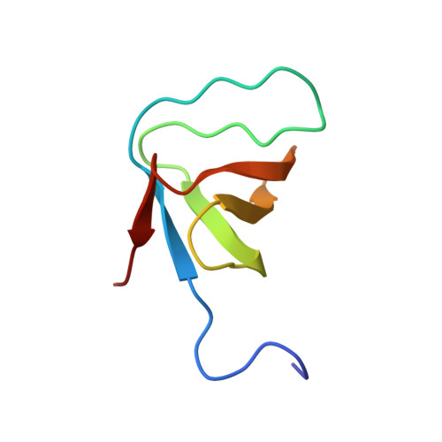
Structural Analysis of the SH3 Domain of beta-PIX and Its Interaction with alpha-p21 Activated Kinase (PAK)

Mott, H.R.Nietlispach, D.Evetts, K.A.Owen, D.

(2005) Biochemistry 44: 10977-10983

  • DOI: https://doi.org/10.1021/bi050374a
  • Primary Citation Related Structures: 
    1ZSG

  • PubMed Abstract: 

    The PAK Ser/Thr kinases are important downstream effectors of the Rho family GTPases Cdc42 and Rac, partly mediating the role of these G proteins in cell proliferation and cytoskeletal rearrangements. As well as small G proteins, PAK interacts with the Cdc42/Rac exchange factor beta-PIX via the PIX SH3 domain and a nontypical Pro-rich region in PAK. This interaction is thought to affect the localization of PAK, as well as increased GTP/GDP exchange of Rac and Cdc42. We have determined the structure of the PIX-SH3/PAK peptide complex and shown that it differs from typical Src-like SH3/peptide complexes. The peptide makes contacts through the Pro-rich sequence in a similar way to standard SH3/peptide complexes, even though the Pro residue positions are not conserved. In addition, there are interactions with a Pro and Lys in the PAK, which are C-terminal to the conserved Arg found in all SH3-binding sequences. These contact a fourth binding pocket on the SH3 domain. We have measured the affinity of PIX-SH3 for the PAK peptide and found that it is of intermediate affinity. When PAK is activated, Ser-199 in the PIX-binding site is phosphorylated. This phosphorylation is sufficient to reduce the affinity for PIX 6-fold.


  • Organizational Affiliation
    • Department of Biochemistry, University of Cambridge, UK. mott@bioc.cam.ac.uk

Macromolecule Content 

  • Total Structure Weight: 9.92 kDa 
  • Atom Count: 701 
  • Modeled Residue Count: 87 
  • Deposited Residue Count: 87 
  • Unique protein chains: 2

Macromolecules

Find similar proteins by:|  3D Structure
Entity ID: 1
MoleculeChains  Sequence LengthOrganismDetailsImage
Rho guanine nucleotide exchange factor 765Homo sapiensMutation(s): 0 
Gene Names: ARHGEF7COOL1KIAA0142P85SPRPAK3BPPIXB
UniProt & NIH Common Fund Data Resources
Find proteins for Q14155 (Homo sapiens)
Explore Q14155 
Go to UniProtKB:  Q14155
PHAROS:  Q14155
GTEx:  ENSG00000102606 
Entity Groups
Sequence Clusters30% Identity50% Identity70% Identity90% Identity95% Identity100% Identity
UniProt GroupQ14155
Sequence Annotations
Expand
Reference Sequence
Find similar proteins by:  Sequence   |   3D Structure  
Entity ID: 2
MoleculeChains  Sequence LengthOrganismDetailsImage
Serine/threonine-protein kinase PAK 122N/AMutation(s): 0 
EC: 2.7.1.37 (PDB Primary Data), 2.7.11.1 (UniProt)
UniProt & NIH Common Fund Data Resources
Find proteins for Q13153 (Homo sapiens)
Explore Q13153 
Go to UniProtKB:  Q13153
PHAROS:  Q13153
GTEx:  ENSG00000149269 
Entity Groups
Sequence Clusters30% Identity50% Identity70% Identity90% Identity95% Identity100% Identity
UniProt GroupQ13153
Sequence Annotations
Expand
Reference Sequence

Experimental Data & Validation

Experimental Data

  • Method: SOLUTION NMR
  • Conformers Calculated: 100 
  • Conformers Submitted: 30 
  • Selection Criteria: structures with the lowest energy 

Structure Validation

View Full Validation Report


Entry History 

Deposition Data

Revision History  (Full details and data files)

  • Version 1.0: 2005-08-30
    Type: Initial release
  • Version 1.1: 2008-04-30
    Changes: Version format compliance
  • Version 1.2: 2011-07-13
    Changes: Version format compliance
  • Version 1.3: 2022-03-02
    Changes: Data collection, Database references, Derived calculations
  • Version 1.4: 2024-05-22
    Changes: Data collection