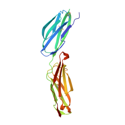
1ZXQ

THE CRYSTAL STRUCTURE OF ICAM-2


Experimental Data Snapshot

  • Method: X-RAY DIFFRACTION
  • Resolution: 2.20 Å
  • R-Value Free: 0.295 
  • R-Value Work: 0.226 
  • R-Value Observed: 0.226 

wwPDB Validation   3D Report Full Report


This is version 2.1 of the entry. See complete history


Literature

Crystal structure of ICAM-2 reveals a distinctive integrin recognition surface.

Casasnovas, J.M.Springer, T.A.Liu, J.H.Harrison, S.C.Wang, J.H.

(1997) Nature 387: 312-315

  • DOI: https://doi.org/10.1038/387312a0
  • Primary Citation of Related Structures:  
    1ZXQ

  • PubMed Abstract: 

    Recognition by integrin proteins on the cell surface regulates the adhesive interactions between cells and their surroundings. The structure of the 'I' domain that is found in some but not all integrins, has been determined. However, the only integrin ligands for which structures are known, namely fibronectin and VCAM-1, are recognized by integrins that lack I domains. The intercellular adhesion molecules ICAM-1, 2 and 3 are, like VCAM-1, members of the immunoglobulin superfamily (IgSF), but they are recognized by an I domain-containing integrin, lymphocyte-function-associated antigen 1 (LFA-1, or CD11a/CD18). Here we present the crystal structure of the extracellular region of ICAM-2. The glutamic acid residue at position 37 is critical for LFA-1 binding and is proposed to coordinate the Mg2+ ion in the I domain; this Glu 37 is surrounded by a relatively flat recognition surface and lies in a beta-strand, whereas the critical aspartic acid residue in VCAM-1 and fibronectin lie in protruding loops. This finding suggests that there are differences in the architecture of recognition sites between integrins that contain or lack I domains. A bend between domains 1 and 2 of ICAM-2 and a tripod-like arrangement of N-linked glycans in the membrane-proximal region of domain 2 may be important for presenting the recognition surface to LFA-1. A model of ICAM-1 based on the ICAM-2 structure provides a framework for understanding its recognition by pathogens.


  • Organizational Affiliation

    The Center for Blood Research, Harvard Medical School, Department of Pathology, Boston, Massachusetts 02115, USA.


Macromolecules
Find similar proteins by:  (by identity cutoff)  |  3D Structure
Entity ID: 1
MoleculeChains Sequence LengthOrganismDetailsImage
INTERCELLULAR ADHESION MOLECULE-2192Homo sapiensMutation(s): 0 
Gene Names: ICAM-2
UniProt & NIH Common Fund Data Resources
Find proteins for P13598 (Homo sapiens)
Explore P13598 
Go to UniProtKB:  P13598
PHAROS:  P13598
GTEx:  ENSG00000108622 
Entity Groups  
Sequence Clusters30% Identity50% Identity70% Identity90% Identity95% Identity100% Identity
UniProt GroupP13598
Glycosylation
Glycosylation Sites: 6Go to GlyGen: P13598-1
Sequence Annotations
Expand
  • Reference Sequence
Oligosaccharides

Help

Entity ID: 2
MoleculeChains Length2D Diagram Glycosylation3D Interactions
2-acetamido-2-deoxy-beta-D-glucopyranose-(1-4)-2-acetamido-2-deoxy-beta-D-glucopyranose
B, C, D, E, F
B, C, D, E, F, G
2N-Glycosylation
Glycosylation Resources
GlyTouCan:  G42666HT
GlyCosmos:  G42666HT
GlyGen:  G42666HT
Experimental Data & Validation

Experimental Data

  • Method: X-RAY DIFFRACTION
  • Resolution: 2.20 Å
  • R-Value Free: 0.295 
  • R-Value Work: 0.226 
  • R-Value Observed: 0.226 
  • Space Group: C 1 2 1
Unit Cell:
Length ( Å )Angle ( ˚ )
a = 69α = 90
b = 63.7β = 114.5
c = 76.2γ = 90
Software Package:
Software NamePurpose
XDSdata scaling
CCP4data reduction
X-PLORmodel building
X-PLORrefinement
XDSdata reduction
CCP4data scaling
X-PLORphasing

Structure Validation

View Full Validation Report


Entry History 

Deposition Data

Revision History  (Full details and data files)

  • Version 1.0: 1997-09-04
    Type: Initial release
  • Version 1.1: 2008-03-24
    Changes: Version format compliance
  • Version 1.2: 2011-07-13
    Changes: Non-polymer description, Version format compliance
  • Version 2.0: 2020-07-29
    Type: Remediation
    Reason: Carbohydrate remediation
    Changes: Atomic model, Data collection, Derived calculations, Structure summary
  • Version 2.1: 2024-10-23
    Changes: Data collection, Database references, Structure summary