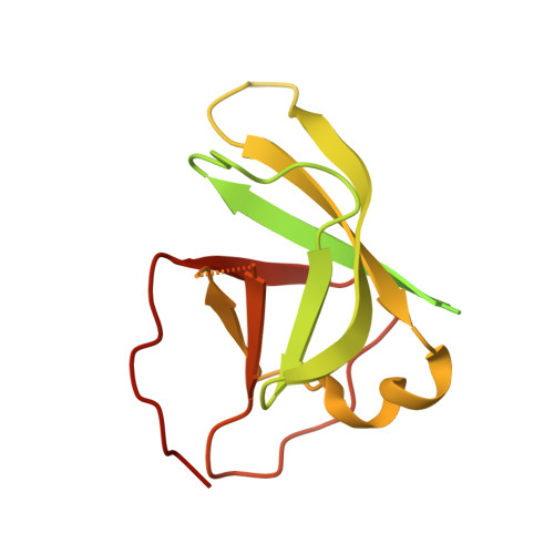
28KO | pdb_000028ko

Cryo-EM structure of B. bacteriovorus Bd1152 2C mutant (I112C, L238C) residues 155-276


Experimental Data Snapshot

  • Method: ELECTRON MICROSCOPY
  • Resolution: 3.40 Å
  • Aggregation State: PARTICLE 
  • Reconstruction Method: SINGLE PARTICLE 

wwPDB Validation 3D Report Full Report

Validation slider image for 28KO

This is version 1.0 of the entry. See complete history. 

Literature

Dynamic ring rearrangements underlie transport by MCE proteins.

Ratkeviciute, G., Cooper, B.F., Iceta-Tena, M., Gragera, M., Caulton, S.G., Wotherspoon, P., Lovering, A.L., Sorzano, C.O.S., Carazo, J.M., Knowles, T.J.

(2026) Structure 

  • DOI: https://doi.org/10.1016/j.str.2026.10.005
  • Primary Citation Related Structures: 
    28KN, 28KO

  • PubMed Abstract: 

    Mammalian cell entry (MCE) proteins mediate hydrophobic substrate transport between bacterial membranes, yet the dynamics underlying substrate translocation remain poorly understood. Here, we present the structural characterization of B. bacteriovorus Bd1152, revealing a rare two-MCE-ring architecture. Integrating X-ray crystallography, cryo-electron microscopy and continuous flexibility analysis, we uncover pronounced shearing and twisting motions between its MCE rings. Analysis of E. coli PqiB reveals that this conformational plasticity extends across distinct MCE architectures, with coordinated inter-ring rearrangements coupled to constriction and dilation of the central transport channel. These dynamics support a peristaltic model for substrate translocation through MCE proteins. Furthermore, a conserved loss-of-function PqiB mutant adopts a collapsed channel conformation, disrupting formation of a continuous transport conduit. Together, our findings establish inter-ring conformational plasticity as a conserved feature of MCE proteins and reveal how coordinated MCE-domain and pore rearrangements may facilitate lipid transport across the bacterial periplasm.


  • Organizational Affiliation: 
    • Institute of Biotechnology, Life Sciences Centre, Vilnius University, 10257 Vilnius, Lithuania. Electronic address: giedre.ratkeviciute-cooper@gmc.vu.lt.

Macromolecule Content 

  • Total Structure Weight: 163.05 kDa 
  • Atom Count: 4,566 
  • Modeled Residue Count: 582 
  • Deposited Residue Count: 1,482 
  • Unique protein chains: 1

Macromolecules

Find similar proteins by:|  3D Structure
Entity ID: 1
MoleculeChains  Sequence LengthOrganismDetailsImage
PqiB protein
A, B, C, D, E
A, B, C, D, E, F
247Bdellovibrio bacteriovorus HD100Mutation(s): 2 
Gene Names: pqiB, Bd1152
UniProt
Find proteins for Q6MNT4 (Bdellovibrio bacteriovorus (strain ATCC 15356 / DSM 50701 / NCIMB 9529 / HD100))
Explore Q6MNT4 
Go to UniProtKB:  Q6MNT4
Entity Groups
Sequence Clusters30% Identity50% Identity70% Identity90% Identity95% Identity100% Identity
UniProt GroupQ6MNT4
Sequence Annotations
Expand
Reference Sequence

Experimental Data & Validation

Experimental Data

  • Method: ELECTRON MICROSCOPY
  • Resolution: 3.40 Å
  • Aggregation State: PARTICLE 
  • Reconstruction Method: SINGLE PARTICLE 
EM Software:
TaskSoftware PackageVersion
MODEL REFINEMENTPHENIX1.21.2_5419
RECONSTRUCTIONcryoSPARC

Structure Validation

View Full Validation Report


Entry History 

& Funding Information

Deposition Data


Funding OrganizationLocationGrant Number
Biotechnology and Biological Sciences Research Council (BBSRC)United KingdomBB/S017283/1
Biotechnology and Biological Sciences Research Council (BBSRC)United KingdomBB/M01116X/1

Revision History  (Full details and data files)

  • Version 1.0: 2026-11-18
    Type: Initial release