2ASU | pdb_00002asu

Crystal Structure of the beta-chain of HGFl/MSP


Experimental Data Snapshot

  • Method: X-RAY DIFFRACTION
  • Resolution: 1.85 Å
  • R-Value Free: 
    0.237 (Depositor), 0.256 (DCC) 
  • R-Value Work: 
    0.198 (Depositor) 
  • R-Value Observed: 
    0.200 (Depositor) 

Starting Model: experimental
View more details

wwPDB Validation 3D Report Full Report

Validation slider image for 2ASU

This is version 1.6 of the entry. See complete history

Literature

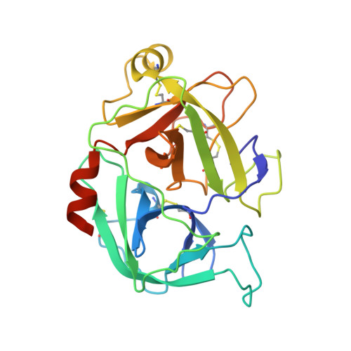
Crystal structure of the beta-chain of human hepatocyte growth factor-like/macrophage stimulating protein.

Carafoli, F.Chirgadze, D.Y.Blundell, T.L.Gherardi, E.

(2005) FEBS J 272: 5799-5807

  • DOI: https://doi.org/10.1111/j.1742-4658.2005.04968.x
  • Primary Citation Related Structures: 
    2ASU

  • PubMed Abstract: 

    Hepatocyte growth factor like/macrophage stimulating protein (HGFl/MSP) and hepatocyte growth factor/scatter factor (HGF/SF) define a distinct family of vertebrate-specific growth factors structurally related to the blood proteinase precursor plasminogen and with important roles in development and cancer. Although the two proteins share a similar domain structure and mechanism of activation, there are differences between HGFl/MSP and HGF/SF in terms of the contribution of individual domains to receptor binding. Here we present a crystal structure of the 30 kDa beta-chain of human HGFl/MSP, a serine proteinase homology domain containing the high-affinity binding site for the RON receptor. The structure describes at 1.85 Angstrom resolution the region of the domain corresponding to the receptor binding site recently defined in the HGF/SF beta-chain, namely the central cleft harboring the three residues corresponding to the catalytic ones of active proteinases (numbers in brackets define the sequence position according to the standard chymotrypsinogen numbering system) [Gln522 (c57), Gln568 (c102) and Tyr661 (c195)] and an adjacent loop flanking the S1 specificity pocket and containing residues Asn682 (c217) and Arg683 (c218) previously shown to be essential for binding of HGFl/MSP to the RON receptor. The study confirms the concept that the serine proteinase homology domains of HGFl/MSP and HGF/SF bind their receptors in an 'enzyme-substrate' mode, reflecting the common evolutionary origin of the plasminogen-related growth factors and the proteinases of the clotting and fibrinolytic pathways. However, analysis of the intermolecular interactions in the crystal lattice of beta-chain HGFl/MSP fails to show the same contacts seen in the HGF/SF structures and does not support a conserved mode of dimerization of the serine proteinase homology domains of HGFl/MSP and HGF/SF responsible for receptor activation.


  • Organizational Affiliation
    • MRC Centre, Hills Road, Cambridge, UK.

Macromolecule Content 

  • Total Structure Weight: 28.35 kDa 
  • Atom Count: 1,884 
  • Modeled Residue Count: 229 
  • Deposited Residue Count: 253 
  • Unique protein chains: 2

Macromolecules

Find similar proteins by:  Sequence   |   3D Structure  
Entity ID: 1
MoleculeChains  Sequence LengthOrganismDetailsImage
Hepatocyte growth factor-like protein19Homo sapiensMutation(s): 0 
Gene Names: MST1HGFL
UniProt & NIH Common Fund Data Resources
Find proteins for P26927 (Homo sapiens)
Explore P26927 
Go to UniProtKB:  P26927
PHAROS:  P26927
Entity Groups
Sequence Clusters30% Identity50% Identity70% Identity90% Identity95% Identity100% Identity
UniProt GroupP26927
Sequence Annotations
Expand
Reference Sequence
Find similar proteins by:|  3D Structure
Entity ID: 2
MoleculeChains  Sequence LengthOrganismDetailsImage
Hepatocyte growth factor-like protein234Homo sapiensMutation(s): 1 
Gene Names: MST1HGFL
UniProt & NIH Common Fund Data Resources
Find proteins for P26927 (Homo sapiens)
Explore P26927 
Go to UniProtKB:  P26927
PHAROS:  P26927
Entity Groups
Sequence Clusters30% Identity50% Identity70% Identity90% Identity95% Identity100% Identity
UniProt GroupP26927
Sequence Annotations
Expand
Reference Sequence

Experimental Data & Validation

Experimental Data

  • Method: X-RAY DIFFRACTION
  • Resolution: 1.85 Å
  • R-Value Free:  0.237 (Depositor), 0.256 (DCC) 
  • R-Value Work:  0.198 (Depositor) 
  • R-Value Observed: 0.200 (Depositor) 
Space Group: P 21 21 21
Unit Cell:
Length ( Å )Angle ( ˚ )
a = 75.676α = 90
b = 75.848β = 90
c = 47.973γ = 90
Software Package:
Software NamePurpose
REFMACrefinement
XDSdata reduction
SCALEPACKdata scaling
AMoREphasing

Structure Validation

View Full Validation Report


Entry History 

Deposition Data

Revision History  (Full details and data files)

  • Version 1.0: 2005-11-10
    Type: Initial release
  • Version 1.1: 2008-04-30
    Changes: Version format compliance
  • Version 1.2: 2011-07-13
    Changes: Version format compliance
  • Version 1.3: 2017-10-11
    Changes: Refinement description
  • Version 1.4: 2021-10-20
    Changes: Database references
  • Version 1.5: 2023-08-23
    Changes: Data collection, Refinement description
  • Version 1.6: 2024-11-06
    Changes: Structure summary